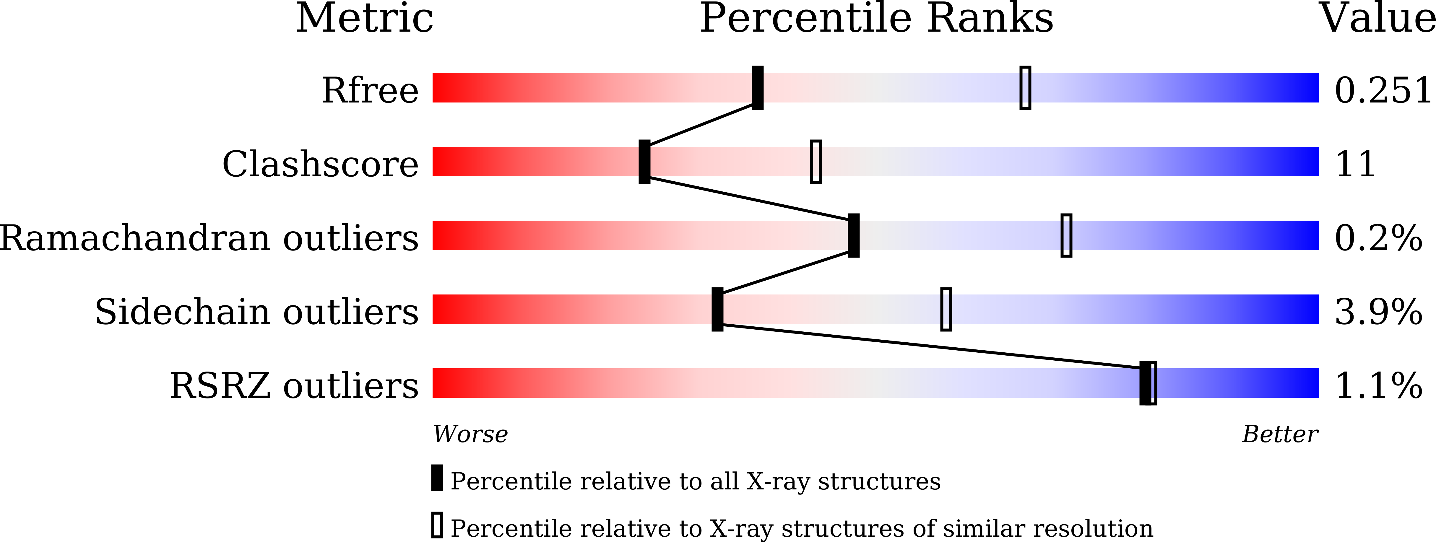
8HD4 image
Deposition Date 2022-11-03
Release Date 2023-02-15
Last Version Date 2024-05-29
Entry Detail
PDB ID:
8HD4
Title:
Full-length crystal structure of mycobacterium tuberculosis FadD23 in complex with AMPC16
Biological Source:
Source Organism:
Method Details:
Experimental Method:
Resolution:
2.68 Å
R-Value Free:
0.25
R-Value Work:
0.21
R-Value Observed:
0.21
Space Group:
P 61 2 2
Macromolecular Entities
Polymer Type:polypeptide(L)
Molecule:Long-chain-fatty-acid--AMP ligase FadD23
Gene (Uniprot):fadD23
Chain IDs:A
Chain Length:605
Number of Molecules:1
Biological Source:Mycobacterium tuberculosis H37Rv
Ligand Molecules
Primary Citation
The Key Roles of Mycobacterium tuberculosis FadD23 C-terminal Domain in Catalytic Mechanisms.
Front Microbiol 14 1090534 1090534 (2023)
PMID: 36896429 DOI: 10.3389/fmicb.2023.1090534

Abstact

Sulfolipid-1 (SL-1) is located in the Mycobacterium tuberculosis (M. tb) cell wall, and is essential for pathogen virulence and intracellular growth. Multiple proteins (e.g., Pks2, FadD23, PapA1, and MmpL8) in the SL-1 synthesis pathway can be treated as drug targets, but, to date, their structures have not been solved. The crystal structures of FadD23 bound to ATP or hexadecanoyl adenylate was determined in this study. We have also investigated long-chain saturated fatty acids as biological substrates of FadD23 through structural, biological, and chemical analyses. The mutation at the active site of FadD23 greatly influences enzymatic activity. Meanwhile, the FadD23 N-terminal domain alone cannot bind palmitic acid without C-terminal domain facilitation since it is almost inactive after removing the C-terminal domain. FadD23 is the first protein in the SL-1 synthesis pathway whose structure has been solved. These results reveal the importance of the C-terminal domain in the catalytic mechanism.

Legend

Protein

Chemical

Disease

Primary Citation of related structures