8HB1 image
Entry Detail
PDB ID:
8HB1
Keywords:
Title:
Crystal structure of NAD-II riboswitch (two strands) with NMN
Biological Source:
PDB Version:
Deposition Date:
2022-10-27
Release Date:
2023-03-22
Method Details:
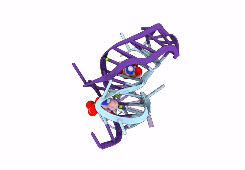
Experimental Method:
Resolution:
2.23 Å
R-Value Free:
0.24
R-Value Work:
0.22
R-Value Observed:
0.22
Space Group:
P 32 2 1
Macromolecular Entities
Polymer Type:polyribonucleotide
Description:RNA (5'-R(*AP*GP*AP*GP*CP*GP*UP*UP*GP*CP*GP*UP*CP*CP*GP*AP*AP*AP*GP*UP*(CBV)P*GP*CP*C)-3')
Chain IDs:A
Chain Length:24
Number of Molecules:1
Biological Source:Streptococcus parasanguinis
Polymer Type:polyribonucleotide
Description:RNA (30-MER)
Chain IDs:B
Chain Length:30
Number of Molecules:1
Biological Source:Streptococcus parasanguinis
Primary Citation
Crystal structures of the NAD+-II riboswitch reveal two distinct ligand-binding pockets.
Nucleic Acids Res. 51 2904 2914 (2023)
PMID: 36840714 DOI: 10.1093/nar/gkad102

Abstact

We present crystal structures of a new NAD+-binding riboswitch termed NAD+-II, bound to nicotinamide mononucleotide (NMN), nicotinamide adenine dinucleotide (NAD+) and nicotinamide riboside (NR). The RNA structure comprises a number of structural features including three helices, one of which forms a triple helix by interacting with an A5 strand in its minor-groove, and another formed from a long-range pseudoknot. The core of the structure (centrally located and coaxial with the triplex and the pseudoknot) includes two consecutive quadruple base interactions. Unusually the riboswitch binds two molecules of ligand, bound at distinct, non-overlapping sites in the RNA. Binding occurs primarily through the nicotinamide moiety of each ligand, held by specific hydrogen bonding and stacking interactions with the pyridyl ring. The mode of binding is the same for NMN, NR and the nicotinamide moiety of NAD+. In addition, when NAD+ is bound into one site it adopts an elongated conformation such that its diphosphate linker occupies a groove on the surface of the RNA, following which the adenine portion inserts into a pocket and makes specific hydrogen bonding interactions. Thus the NAD+-II riboswitch is distinct from the NAD+-I riboswitch in that it binds two molecules of ligand at separate sites, and that binding occurs principally through the nicotinamide moiety.

Legend

Protein

Chemical

Disease

Primary Citation of related structures