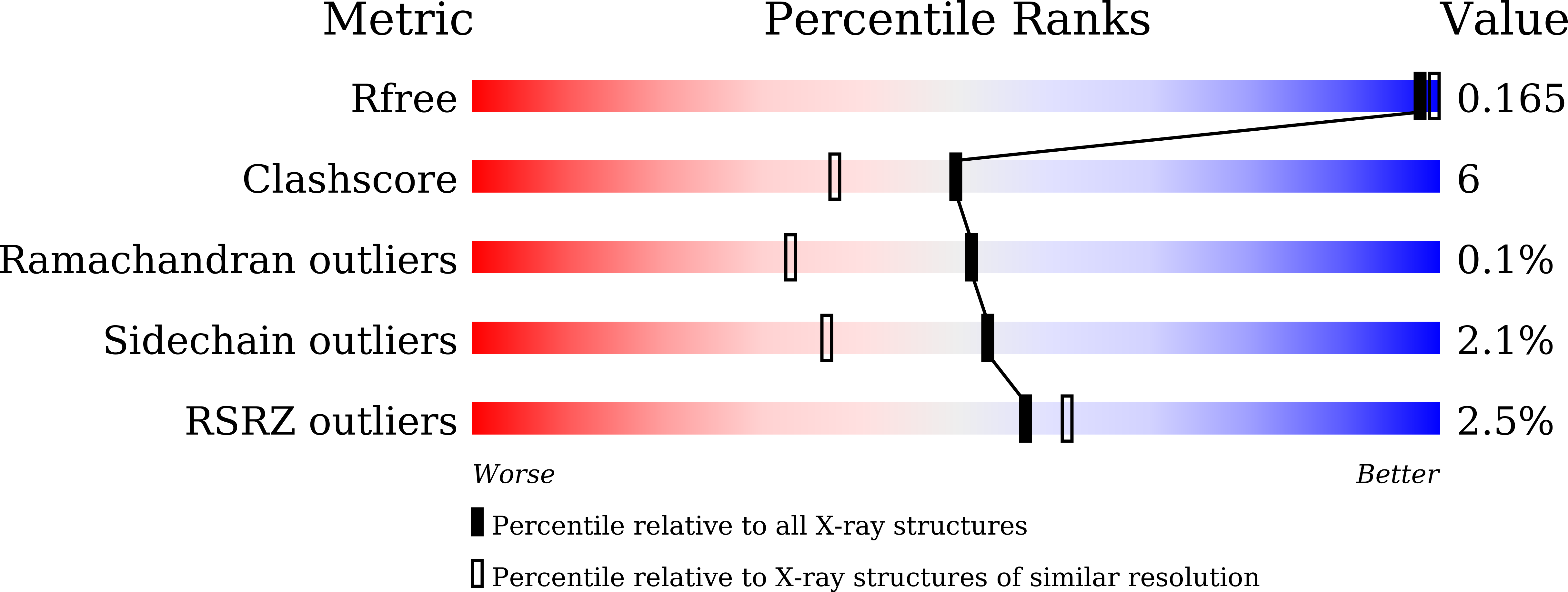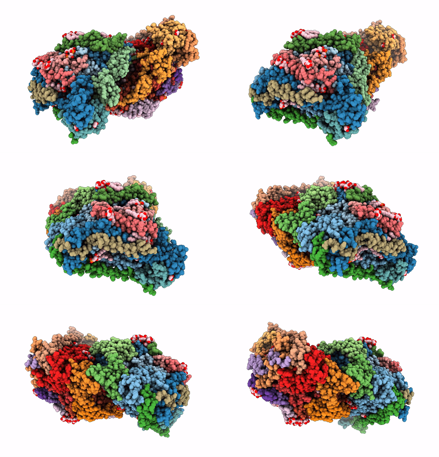
8H8R image
Deposition Date 2022-10-24
Release Date 2023-02-08
Last Version Date 2024-11-06
Entry Detail
PDB ID:
8H8R
Keywords:
Title:
Bovine Heart Cytochrome c Oxidase in the Calcium-bound Fully Oxidized State
Biological Source:
Source Organism:
Bos taurus (Taxon ID: 9913)
Host Organism:
Method Details:
Experimental Method:
Resolution:
1.70 Å
R-Value Free:
0.15
R-Value Work:
0.12
R-Value Observed:
0.12
Space Group:
P 21 21 21
Macromolecular Entities
Polymer Type:polypeptide(L)
Molecule:Cytochrome c oxidase subunit 1
Gene (Uniprot):MT-CO1
Chain IDs:A, N
Chain Length:514
Number of Molecules:2
Biological Source:Bos taurus
Polymer Type:polypeptide(L)
Molecule:Cytochrome c oxidase subunit 2
Gene (Uniprot):MT-CO2
Chain IDs:B, O
Chain Length:227
Number of Molecules:2
Biological Source:Bos taurus
Polymer Type:polypeptide(L)
Molecule:Cytochrome c oxidase subunit 3
Gene (Uniprot):MT-CO3
Chain IDs:C, P
Chain Length:261
Number of Molecules:2
Biological Source:Bos taurus
Polymer Type:polypeptide(L)
Molecule:Cytochrome c oxidase subunit 4 isoform 1, mitochondrial
Gene (Uniprot):COX4I1
Chain IDs:D, Q
Chain Length:147
Number of Molecules:2
Biological Source:Bos taurus
Polymer Type:polypeptide(L)
Molecule:Cytochrome c oxidase subunit 5A
Gene (Uniprot):COX5A
Chain IDs:E, R
Chain Length:109
Number of Molecules:2
Biological Source:Bos taurus
Polymer Type:polypeptide(L)
Molecule:Cytochrome c oxidase subunit 5B, mitochondrial
Gene (Uniprot):COX5B
Chain IDs:F, S
Chain Length:98
Number of Molecules:2
Biological Source:Bos taurus
Polymer Type:polypeptide(L)
Molecule:Cytochrome c oxidase subunit 6A2, mitochondrial
Gene (Uniprot):COX6A2
Chain IDs:G, T
Chain Length:85
Number of Molecules:2
Biological Source:Bos taurus
Polymer Type:polypeptide(L)
Molecule:Cytochrome c oxidase subunit 6B1
Gene (Uniprot):COX6B1
Chain IDs:H, U
Chain Length:85
Number of Molecules:2
Biological Source:Bos taurus
Polymer Type:polypeptide(L)
Molecule:Cytochrome c oxidase subunit 6C
Gene (Uniprot):COX6C
Chain IDs:I, V
Chain Length:73
Number of Molecules:2
Biological Source:Bos taurus
Polymer Type:polypeptide(L)
Molecule:Cytochrome c oxidase subunit 7A1
Gene (Uniprot):COX7A1
Chain IDs:J, W
Chain Length:59
Number of Molecules:2
Biological Source:Bos taurus
Polymer Type:polypeptide(L)
Molecule:Cytochrome c oxidase subunit 7B, mitochondrial
Gene (Uniprot):COX7B
Chain IDs:K, X
Chain Length:56
Number of Molecules:2
Biological Source:Bos taurus
Polymer Type:polypeptide(L)
Molecule:Cytochrome c oxidase subunit 7C, mitochondrial
Gene (Uniprot):COX7C
Chain IDs:L, Y
Chain Length:47
Number of Molecules:2
Biological Source:Bos taurus
Polymer Type:polypeptide(L)
Molecule:Cytochrome c oxidase subunit 8B, mitochondrial
Gene (Uniprot):COX8B
Chain IDs:M, Z
Chain Length:46
Number of Molecules:2
Biological Source:Bos taurus
Modified Residue
Compound ID Chain ID Parent Comp ID Details 2D Image
FME A MET modified residue
Primary Citation
Calcium-bound structure of bovine cytochrome c oxidase.
Biochim Biophys Acta Bioenerg 1864 148956 148956 (2023)
PMID: 36708913 DOI: 10.1016/j.bbabio.2023.148956

Abstact

The crystal structure of bovine cytochrome c oxidase (CcO) shows a sodium ion (Na+) bound to the surface of subunit I. Changes in the absorption spectrum of heme a caused by calcium ions (Ca2+) are detected as small red shifts, and inhibition of enzymatic activity under low turnover conditions is observed by addition of Ca2+ in a competitive manner with Na+. In this study, we determined the crystal structure of Ca2+-bound bovine CcO in the oxidized and reduced states at 1.7 Å resolution. Although Ca2+ and Na+ bound to the same site of oxidized and reduced CcO, they led to different coordination geometries. Replacement of Na+ with Ca2+ caused a small structural change in the loop segments near the heme a propionate and formyl groups, resulting in spectral changes in heme a. Redox-coupled structural changes observed in the Ca2+-bound form were the same as those previously observed in the Na+-bound form, suggesting that binding of Ca2+ does not severely affect enzymatic function, which depends on these structural changes. The relation between the Ca2+ binding and the inhibitory effect during slow turnover, as well as the possible role of bound Ca2+ are discussed.

Legend

Protein

Chemical

Disease

Primary Citation of related structures