
Deposition Date
2022-10-19
Release Date
2023-09-20
Last Version Date
2023-09-20
Entry Detail
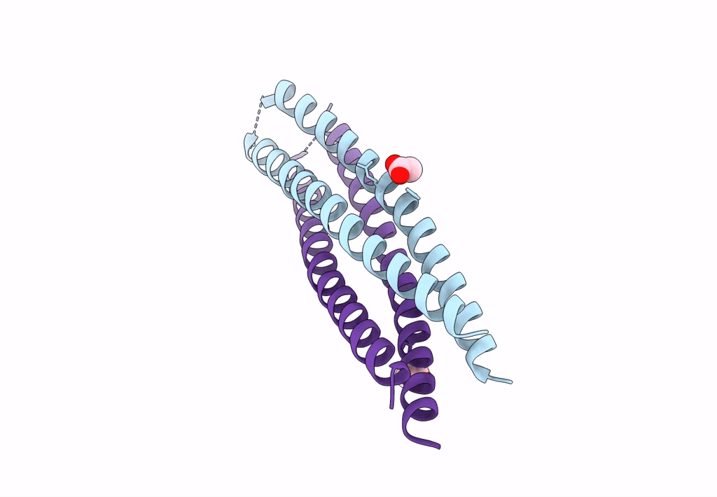
PDB ID:
8H7D
Keywords:
Title:
Crystal structure of a de novo enzyme, ferric enterobactin esterase Syn-F4 (K4T)
Biological Source:
Source Organism(s):
synthetic construct (Taxon ID: 32630)
Expression System(s):
Method Details:
Experimental Method:
Resolution:
2.20 Å
R-Value Free:
0.30
R-Value Work:
0.24
R-Value Observed:
0.24
Space Group:
C 2 2 21
