Deposition Date
2022-10-18
Release Date
2023-08-02
Last Version Date
2024-10-23
Entry Detail
PDB ID:
8H73
Keywords:
Title:
Crystal structure of antibody scFv against M2e Influenza peptide
Biological Source:
Source Organism(s):
Homo sapiens (Taxon ID: 9606)
Expression System(s):
Method Details:
Experimental Method:
Resolution:
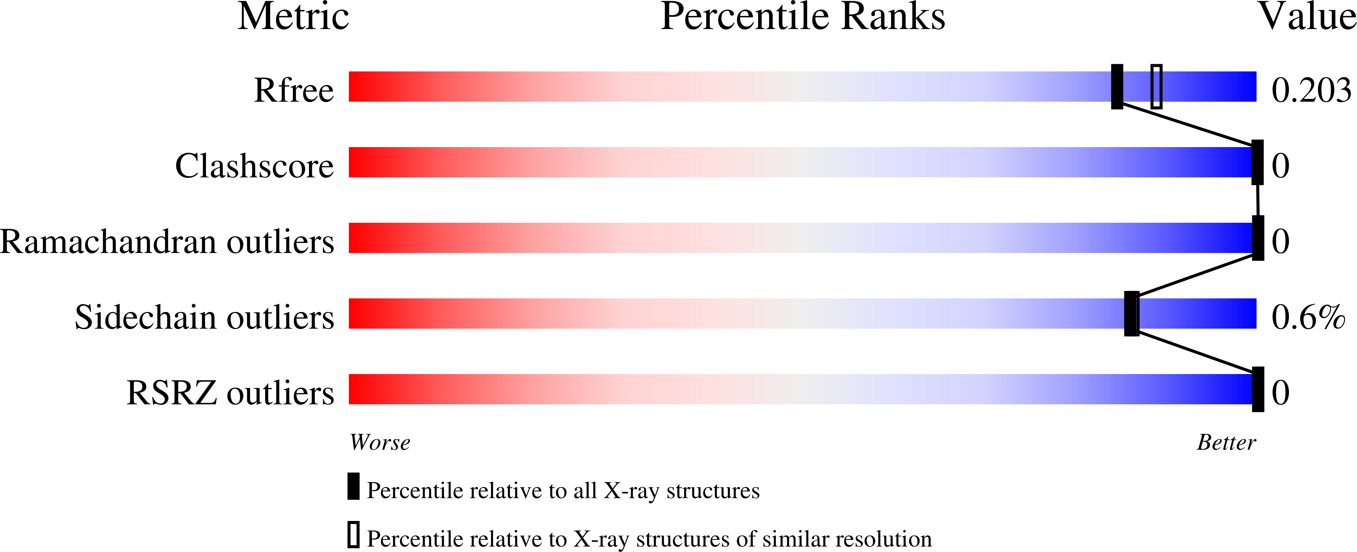
1.91 Å
R-Value Free:
0.20
R-Value Work:
0.18
R-Value Observed:
0.18
Space Group:
P 61 2 2