8H2C image
Deposition Date 2022-10-05
Release Date 2022-11-09
Last Version Date 2024-04-03
Entry Detail
PDB ID:
8H2C
Title:
Crystal structure of the pseudaminic acid synthase PseI from Campylobacter jejuni
Biological Source:
Source Organism:
Method Details:
Experimental Method:
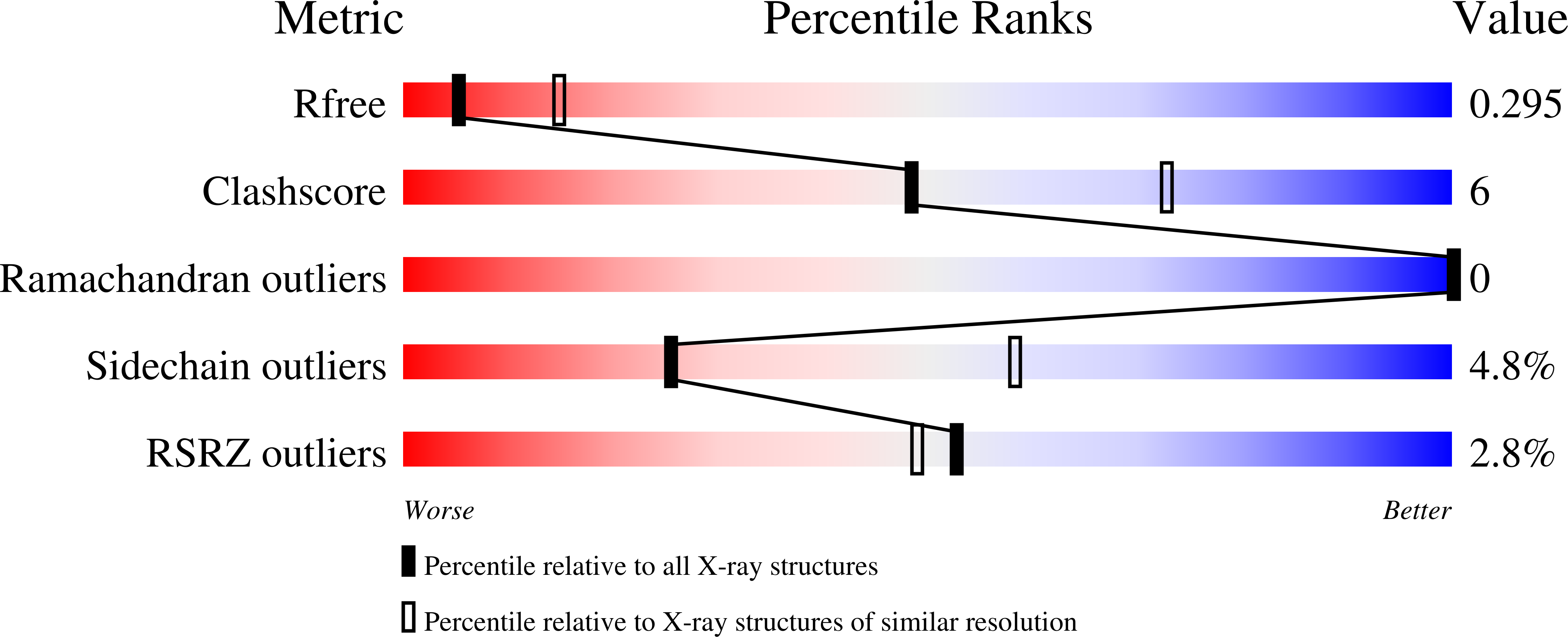
Resolution:
2.90 Å
R-Value Free:
0.29
R-Value Work:
0.24
R-Value Observed:
0.24
Space Group:
P 4 21 2
Macromolecular Entities
Polymer Type:polypeptide(L)
Molecule:Pseudaminic acid synthase
Chain IDs:A, B, C, D
Chain Length:349
Number of Molecules:4
Biological Source:Campylobacter jejuni
Ligand Molecules
Primary Citation
Structural analysis of the pseudaminic acid synthase PseI from Campylobacter jejuni.
Biochem.Biophys.Res.Commun. 635 252 258 (2022)
PMID: 36283338 DOI: 10.1016/j.bbrc.2022.10.050

Abstact

Campylobacter jejuni PseI is a pseudaminic acid synthase that condenses the 2,4-diacetamido-2,4,6-trideoxy-l-altrose sugar (6-deoxy AltdiNAc) and phosphoenolpyruvate to generate pseudaminic acid, a sialic acid-like 9-carbon backbone α-keto sugar. Pseudaminic acid is conjugated to cell surface proteins and lipids and plays a key role in the mobility and virulence of C. jejuni and other pathogenic bacteria. To provide insights into the catalytic mechanism of PseI, we performed a structural study on PseI. PseI forms a two-domain structure and assembles into a domain-swapped homodimer. The PseI dimer has two cavities, each of which accommodates a metal ion using conserved histidine residues. A comparative analysis of structures and sequences suggests that the cavity of PseI functions as an active site that binds the 6-deoxy AltdiNAc and phosphoenolpyruvate substrates and mediates their condensation. Furthermore, we propose the substrate binding-induced structural rearrangement of PseI and predict 6-deoxy AltdiNAc recognition residues that are specific to PseI.

Legend

Protein

Chemical

Disease

Primary Citation of related structures