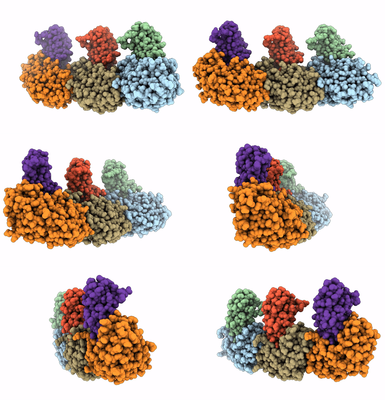
8GZW image
Deposition Date 2022-09-27
Release Date 2023-07-19
Last Version Date 2023-11-29
Entry Detail
PDB ID:
8GZW
Keywords:
Title:
Klebsiella pneumoniae FtsZ complexed with monobody (P21)
Biological Source:
Source Organism:
Host Organism:
Method Details:
Experimental Method:
Resolution:
2.50 Å
R-Value Free:
0.28
R-Value Work:
0.21
R-Value Observed:
0.21
Space Group:
P 1 21 1
Macromolecular Entities
Polymer Type:polypeptide(L)
Molecule:Cell division protein FtsZ
Chain IDs:A, C, E
Chain Length:309
Number of Molecules:3
Biological Source:Klebsiella pneumoniae
Polymer Type:polypeptide(L)
Molecule:Monobody
Chain IDs:B, D, F
Chain Length:92
Number of Molecules:3
Biological Source:Homo sapiens
Ligand Molecules
Primary Citation
Structures of a FtsZ single protofilament and a double-helical tube in complex with a monobody.
Nat Commun 14 4073 4073 (2023)
PMID: 37429870 DOI: 10.1038/s41467-023-39807-5

Abstact

FtsZ polymerizes into protofilaments to form the Z-ring that acts as a scaffold for accessory proteins during cell division. Structures of FtsZ have been previously solved, but detailed mechanistic insights are lacking. Here, we determine the cryoEM structure of a single protofilament of FtsZ from Klebsiella pneumoniae (KpFtsZ) in a polymerization-preferred conformation. We also develop a monobody (Mb) that binds to KpFtsZ and FtsZ from Escherichia coli without affecting their GTPase activity. Crystal structures of the FtsZ-Mb complexes reveal the Mb binding mode, while addition of Mb in vivo inhibits cell division. A cryoEM structure of a double-helical tube of KpFtsZ-Mb at 2.7 Å resolution shows two parallel protofilaments. Our present study highlights the physiological roles of the conformational changes of FtsZ in treadmilling that regulate cell division.

Legend

Protein

Chemical

Disease

Primary Citation of related structures