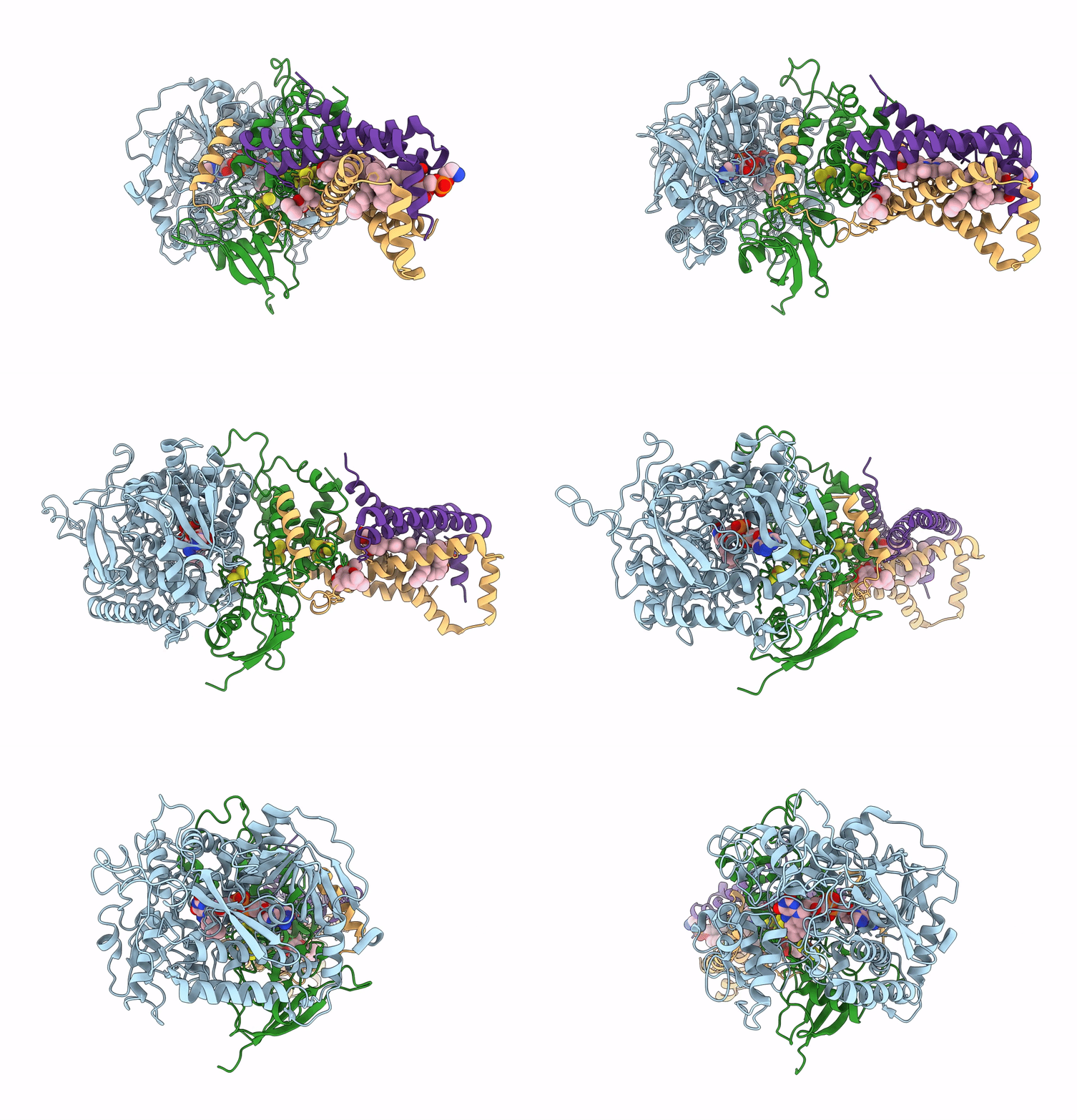
8GS8 image
Deposition Date 2022-09-05
Release Date 2023-05-10
Last Version Date 2025-07-02
Entry Detail
PDB ID:
8GS8
Keywords:
Title:
cryo-EM structure of the human respiratory complex II
Biological Source:
Source Organism:
Homo sapiens (Taxon ID: 9606)
Host Organism:
Method Details:
Experimental Method:
Resolution:
2.86 Å
Aggregation State:
PARTICLE
Reconstruction Method:
SINGLE PARTICLE
Macromolecular Entities
Polymer Type:polypeptide(L)
Molecule:Succinate dehydrogenase [ubiquinone] flavoprotein subunit, mitochondrial
Gene (Uniprot):SDHA
Chain IDs:A
Chain Length:664
Number of Molecules:1
Biological Source:Homo sapiens
Polymer Type:polypeptide(L)
Molecule:Succinate dehydrogenase [ubiquinone] iron-sulfur subunit, mitochondrial
Gene (Uniprot):SDHB
Chain IDs:B
Chain Length:280
Number of Molecules:1
Biological Source:Homo sapiens
Polymer Type:polypeptide(L)
Molecule:Succinate dehydrogenase cytochrome b560 subunit, mitochondrial
Gene (Uniprot):SDHC
Chain IDs:C
Chain Length:169
Number of Molecules:1
Biological Source:Homo sapiens
Polymer Type:polypeptide(L)
Molecule:Succinate dehydrogenase [ubiquinone] cytochrome b small subunit, mitochondrial
Gene (Uniprot):SDHD
Chain IDs:D
Chain Length:159
Number of Molecules:1
Biological Source:Homo sapiens
Primary Citation
Structure of the human respiratory complex II.
Proc.Natl.Acad.Sci.USA 120 e2216713120 e2216713120 (2023)
PMID: 37098072 DOI: 10.1073/pnas.2216713120

Abstact

Human complex II is a key protein complex that links two essential energy-producing processes: the tricarboxylic acid cycle and oxidative phosphorylation. Deficiencies due to mutagenesis have been shown to cause mitochondrial disease and some types of cancers. However, the structure of this complex is yet to be resolved, hindering a comprehensive understanding of the functional aspects of this molecular machine. Here, we have determined the structure of human complex II in the presence of ubiquinone at 2.86 Å resolution by cryoelectron microscopy, showing it comprises two water-soluble subunits, SDHA and SDHB, and two membrane-spanning subunits, SDHC and SDHD. This structure allows us to propose a route for electron transfer. In addition, clinically relevant mutations are mapped onto the structure. This mapping provides a molecular understanding to explain why these variants have the potential to produce disease.

Legend

Protein

Chemical

Disease

Primary Citation of related structures