8GR2 image
Deposition Date 2022-08-31
Release Date 2022-11-16
Last Version Date 2024-04-03
Entry Detail
PDB ID:
8GR2
Keywords:
Title:
Crystal structure of the GDSL-family esterase CJ0610C from Campylobacter jejuni
Biological Source:
Source Organism(s):
Expression System(s):
Method Details:
Experimental Method:
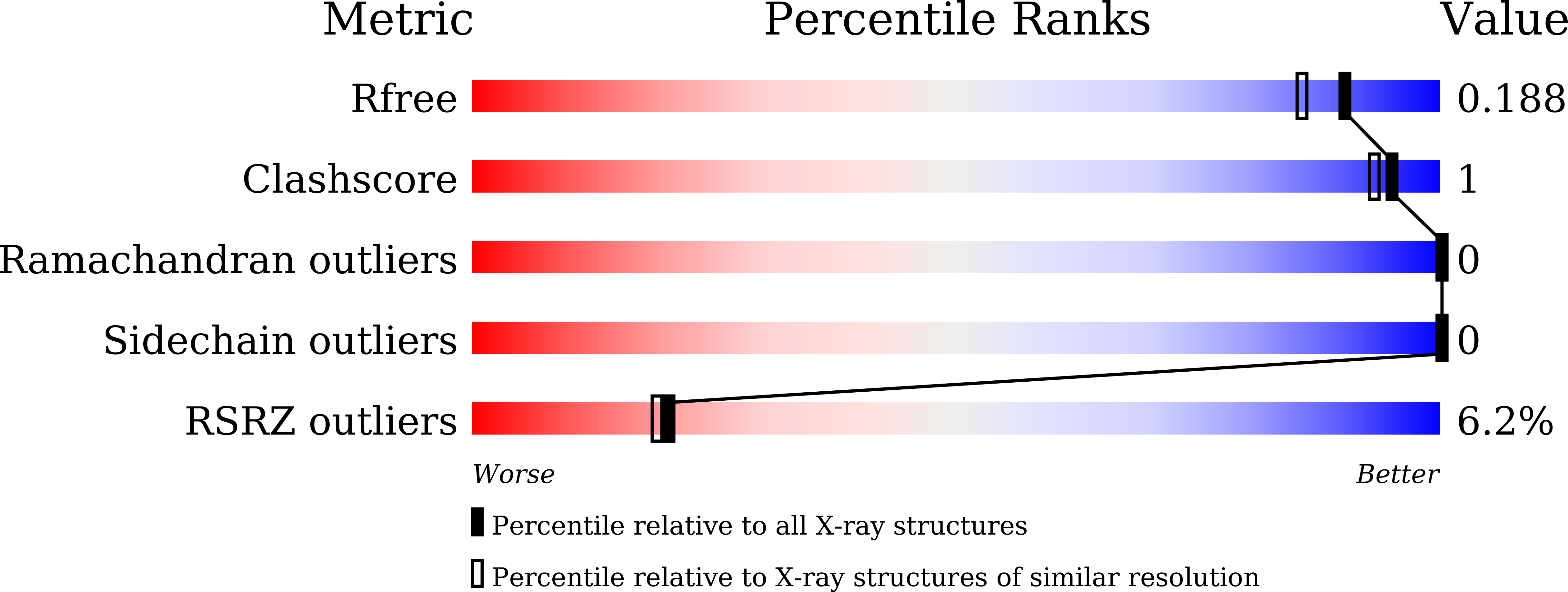
Resolution:
1.65 Å
R-Value Free:
0.18
R-Value Work:
0.16
R-Value Observed:
0.16
Space Group:
P 61
Macromolecular Entities
Polymer Type:polypeptide(L)
Molecule:DUF459 domain-containing protein
Chain IDs:A
Chain Length:227
Number of Molecules:1
Biological Source:Campylobacter jejuni
Ligand Molecules
Primary Citation
Structural and biochemical analysis of the GDSL-family esterase CJ0610C from Campylobacter jejuni.
Biochem.Biophys.Res.Commun. 631 124 129 (2022)
PMID: 36183553 DOI: 10.1016/j.bbrc.2022.09.071

Abstact

GDSL domain-containing proteins generally hydrolyze esters or lipids and play critical roles in diverse biological and industrial processes. GDSL hydrolases use catalytic triad and oxyanion hole residues from conserved blocks I, II, III, and V to drive the esterase reaction. However, GDSL hydrolases exhibit large deviations in sequence, structure, and substrate specificity, requiring the characterization of each GDSL hydrolase to reveal its catalytic mechanism. We identified a GDSL protein (CJ0610C) from pathogenic Campylobacter jejuni and assessed its biochemical and structural features. CJ0610C displayed esterase activity for p-nitrophenyl acetate and preferred short chain esters and alkaline pH. The C-terminal two-thirds of CJ0610C corresponding to the GDSL domain forms a three-layered α/β/α fold as a core structure in which a five-stranded β-sheet is sandwiched by α-helices. In the CJ0610C structure, conserved catalytic triad and oxyanion hole residues that are indispensable for esterase activity are found in blocks I, III, and V. However, CJ0610C lacks the conserved block-II glycine residue and instead employs a unique asparagine residue as another oxyanion hole residue. Moreover, our structural analysis suggests that substrate binding is mediated by a CJ0610C-specific pocket, which is surrounded by hydrophobic residues and occluded at one end by a positively charged arginine residue.

Legend

Protein

Chemical

Disease

Primary Citation of related structures
Feedback Form
Name
Email
Institute
Feedback