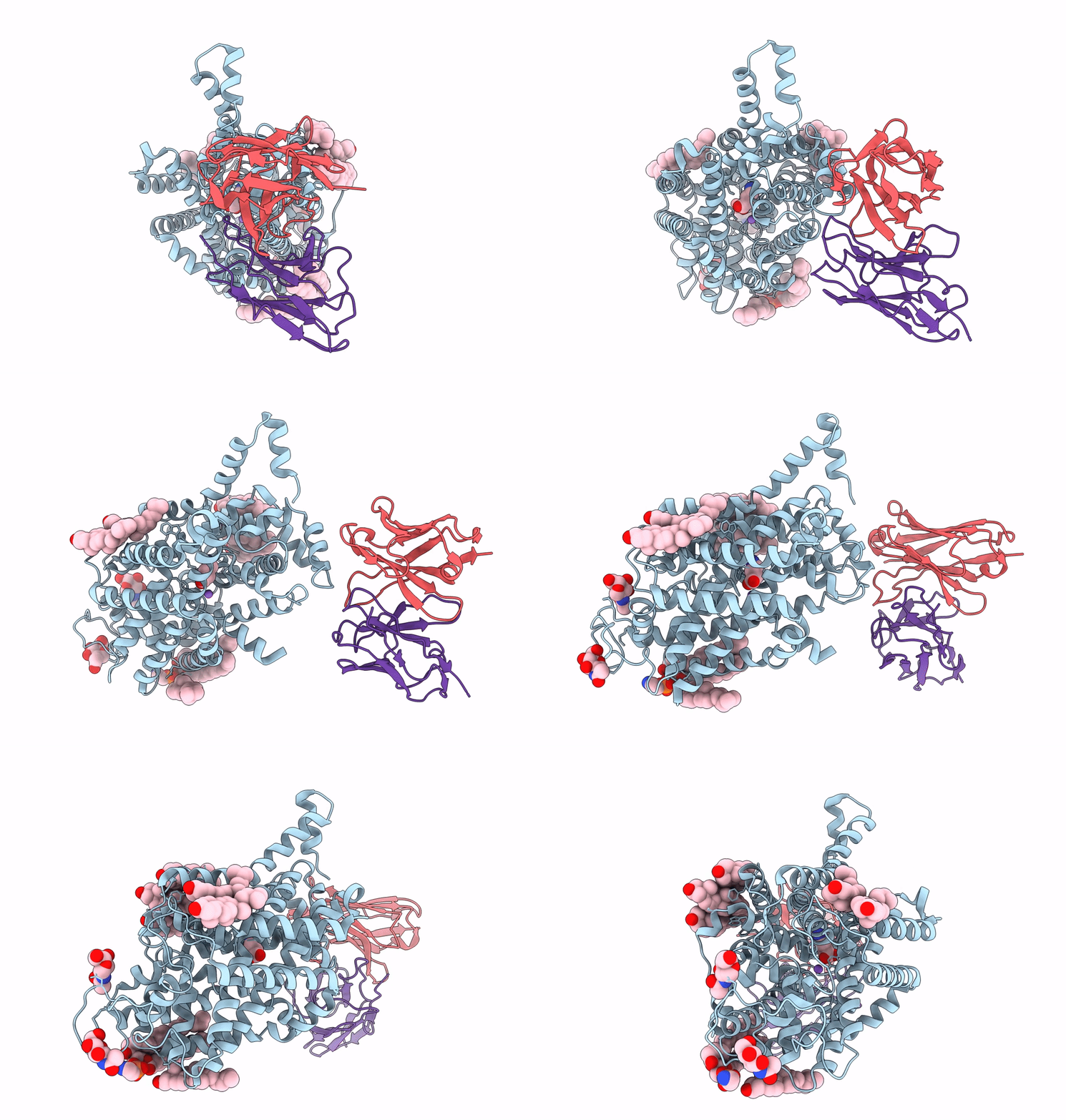
8GNK image
Deposition Date 2022-08-24
Release Date 2023-05-31
Last Version Date 2024-10-23
Entry Detail
PDB ID:
8GNK
Title:
CryoEM structure of cytosol-facing, substrate-bound ratGAT1
Biological Source:
Source Organism:
Rattus norvegicus (Taxon ID: 10116)
Mus musculus (Taxon ID: 10090)
Host Organism:
Method Details:
Experimental Method:
Resolution:
3.10 Å
Aggregation State:
PARTICLE
Reconstruction Method:
SINGLE PARTICLE
Macromolecular Entities
Structures with similar UniProt ID
Protein Blast
Polymer Type:polypeptide(L)
Molecule:Sodium- and chloride-dependent GABA transporter 1
Gene (Uniprot):Slc6a1
Mutagens:F312Y, Y481S, D482E, N483D, Q485R, E486D, V488I, S490F, R491P
Chain IDs:A
Chain Length:562
Number of Molecules:1
Biological Source:Rattus norvegicus
Polymer Type:polypeptide(L)
Molecule:fragment antigen binding 9D5 heavy chain
Chain IDs:C (auth: H)
Chain Length:219
Number of Molecules:1
Biological Source:Mus musculus
Polymer Type:polypeptide(L)
Molecule:Fragment antigen binding light chain
Chain IDs:B (auth: L)
Chain Length:214
Number of Molecules:1
Biological Source:Mus musculus
Primary Citation
Cryo-EM structure of GABA transporter 1 reveals substrate recognition and transport mechanism.
Nat.Struct.Mol.Biol. 30 1023 1032 (2023)
PMID: 37400654 DOI: 10.1038/s41594-023-01011-w

Abstact

The inhibitory neurotransmitter γ-aminobutyric acid (GABA) is cleared from the synaptic cleft by the sodium- and chloride-coupled GABA transporter GAT1. Inhibition of GAT1 prolongs the GABAergic signaling at the synapse and is a strategy to treat certain forms of epilepsy. In this study, we present the cryo-electron microscopy structure of Rattus norvegicus GABA transporter 1 (rGAT1) at a resolution of 3.1 Å. The structure elucidation was facilitated by epitope transfer of a fragment-antigen binding (Fab) interaction site from the Drosophila dopamine transporter (dDAT) to rGAT1. The structure reveals rGAT1 in a cytosol-facing conformation, with a linear density in the primary binding site that accommodates a molecule of GABA, a displaced ion density proximal to Na site 1 and a bound chloride ion. A unique insertion in TM10 aids the formation of a compact, closed extracellular gate. Besides yielding mechanistic insights into ion and substrate recognition, our study will enable the rational design of specific antiepileptics.

Legend

Protein

Chemical

Disease

Primary Citation of related structures