Deposition Date
2023-03-22
Release Date
2023-09-20
Last Version Date
2024-10-16
Entry Detail
PDB ID:
8GLG
Keywords:
Title:
Crystal Structure of Human CD1b in Complex with Phosphatidylethanolamine C34:1
Biological Source:
Source Organism(s):
Homo sapiens (Taxon ID: 9606)
Expression System(s):
Method Details:
Experimental Method:
Resolution:
1.60 Å
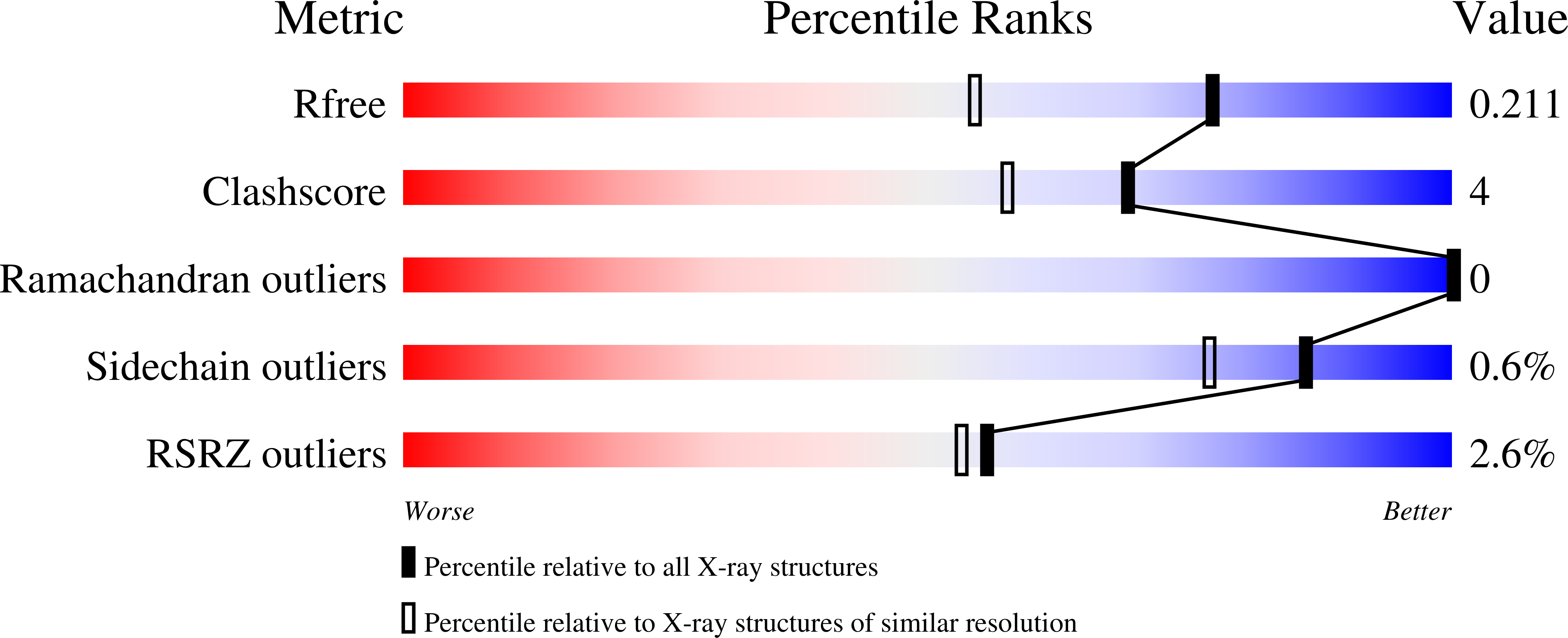
R-Value Free:
0.20
R-Value Work:
0.18
R-Value Observed:
0.18
Space Group:
P 21 21 21