Deposition Date
2023-03-02
Release Date
2023-09-27
Last Version Date
2023-09-27
Entry Detail
PDB ID:
8GCQ
Keywords:
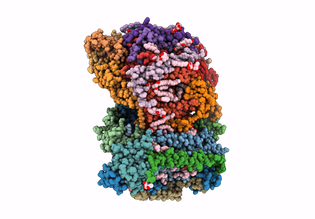
Title:
SFX structure of oxidized cytochrome c oxidase at 2.38 Angstrom resolution
Biological Source:
Source Organism(s):
Bos taurus (Taxon ID: 9913)
Method Details:
Experimental Method:
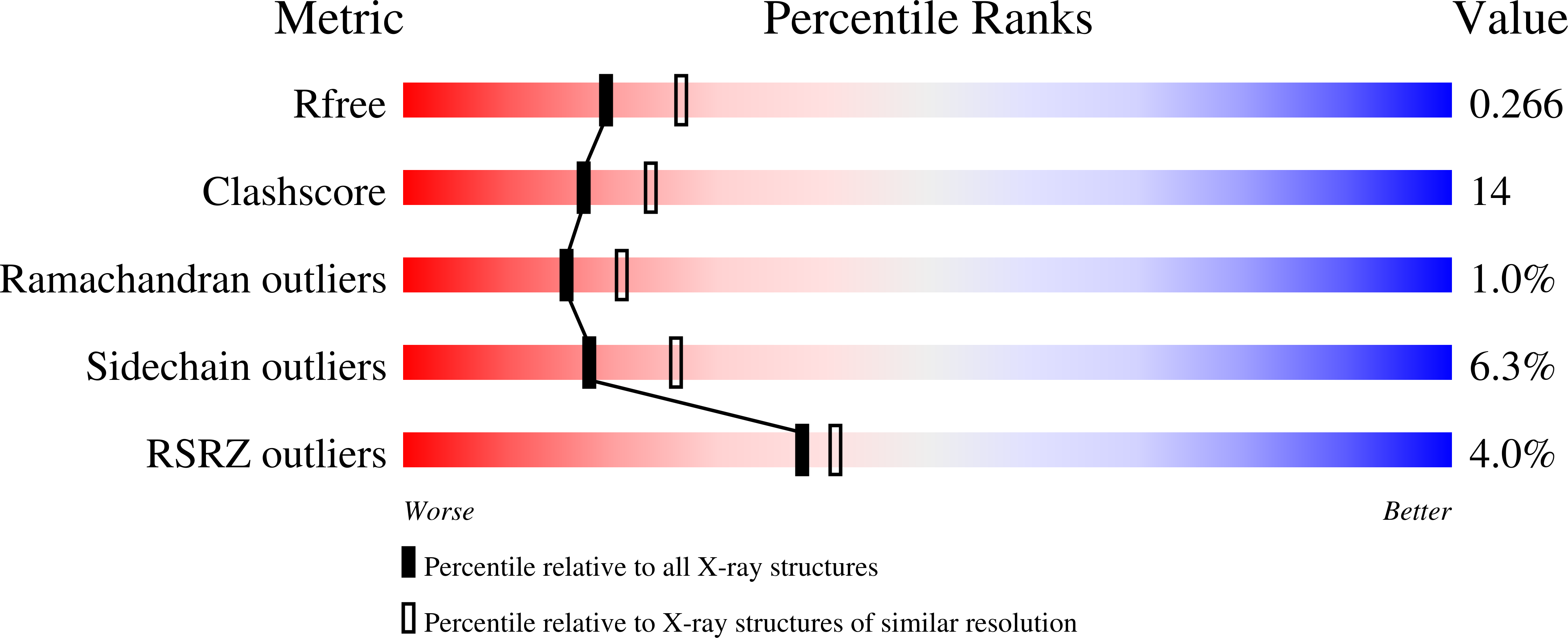
Resolution:
2.38 Å
R-Value Free:
0.26
R-Value Work:
0.23
Space Group:
P 21 21 21