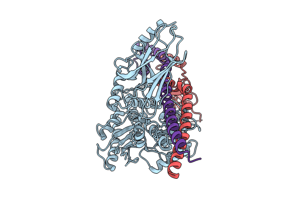
8GB3 image
Deposition Date 2023-02-24
Release Date 2024-01-31
Last Version Date 2024-05-01
Entry Detail
PDB ID:
8GB3
Keywords:
Title:
Structure of the Mycobacterium tuberculosis Hsp70 protein DnaK bound to the nucleotide exchange factor GrpE
Biological Source:
Source Organism:
Host Organism:
Method Details:
Experimental Method:
Resolution:
3.70 Å
Aggregation State:
PARTICLE
Reconstruction Method:
SINGLE PARTICLE
Macromolecular Entities
Polymer Type:polypeptide(L)
Molecule:Chaperone protein DnaK
Chain IDs:A (auth: D)
Chain Length:625
Number of Molecules:1
Biological Source:Mycobacterium tuberculosis
Polymer Type:polypeptide(L)
Molecule:Protein GrpE
Chain IDs:B (auth: E), C (auth: F)
Chain Length:235
Number of Molecules:2
Biological Source:Mycobacterium tuberculosis
Primary Citation
Structure of the M. tuberculosis DnaK-GrpE complex reveals how key DnaK roles are controlled.
Nat Commun 15 660 660 (2024)
PMID: 38253530 DOI: 10.1038/s41467-024-44933-9

Abstact

The molecular chaperone DnaK is essential for viability of Mycobacterium tuberculosis (Mtb). DnaK hydrolyzes ATP to fold substrates, and the resulting ADP is exchanged for ATP by the nucleotide exchange factor GrpE. It has been unclear how GrpE couples DnaK's nucleotide exchange with substrate release. Here we report a cryo-EM analysis of GrpE bound to an intact Mtb DnaK, revealing an asymmetric 1:2 DnaK-GrpE complex. The GrpE dimer ratchets to modulate both DnaK nucleotide-binding domain and the substrate-binding domain. We further show that the disordered GrpE N-terminus is critical for substrate release, and that the DnaK-GrpE interface is essential for protein folding activity both in vitro and in vivo. Therefore, the Mtb GrpE dimer allosterically regulates DnaK to concomitantly release ADP in the nucleotide-binding domain and substrate peptide in the substrate-binding domain.

Legend

Protein

Chemical

Disease

Primary Citation of related structures