
Deposition Date
2023-02-21
Release Date
2024-02-21
Last Version Date
2024-09-18
Entry Detail
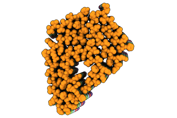
PDB ID:
8G9R
Keywords:
Title:
Cardiac amyloid fibrils extracted from a wild-type ATTR amyloidosis patient
Biological Source:
Source Organism(s):
Homo sapiens (Taxon ID: 9606)
Method Details:
Experimental Method:
Resolution:
3.28 Å
Aggregation State:
HELICAL ARRAY
Reconstruction Method:
HELICAL
