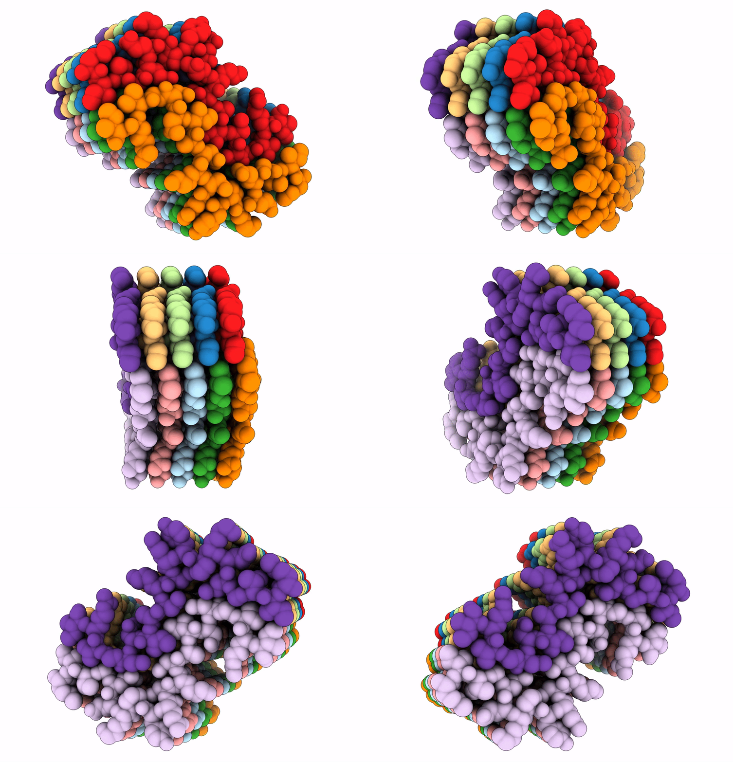
8G2V image
Entry Detail
PDB ID:
8G2V
EMDB ID:
Keywords:
Title:
Cryo-EM structure of recombinant human LECT2 amyloid fibril core
Biological Source:
Source Organism:
Host Organism:
PDB Version:
Deposition Date:
2023-02-06
Release Date:
2023-08-16
Method Details:
Experimental Method:
Resolution:
2.72 Å
Aggregation State:
FILAMENT
Reconstruction Method:
HELICAL
Macromolecular Entities
Polymer Type:polypeptide(L)
Description:Leukocyte cell-derived chemotaxin-2
Chain IDs:A, B, C, D, E, F, G, H, I, J
Chain Length:21
Number of Molecules:10
Biological Source:Homo sapiens
Ligand Molecules
Primary Citation
Cryo-EM structure of a human LECT2 amyloid fibril reveals a network of polar ladders at its core.
Structure 31 1386 1393.e3 (2023)
PMID: 37657439 DOI: 10.1016/j.str.2023.08.007

Abstact

ALECT2 systemic amyloidosis is associated with deposition of the leukocyte cell-derived chemotaxin-2 (LECT2) protein in the form of fibrils. In ALECT2 amyloidosis, ALECT2 fibrils deposit in the glomerulus, resulting in renal failure. Patients lack effective treatment options outside of renal transplant or dialysis. The structure of globular LECT2 has been determined but structures of ALECT2 amyloid fibrils remain unknown. Using single-particle cryo-EM, we find that recombinant human LECT2 forms robust twisting fibrils with canonical amyloid features. ALECT2 fibrils contain two mating protofilaments spanning residues 55-75 of the LECT2 sequence. The geometry of the ALECT2 fibril displays features in line with other pathogenic amyloids. Its core is tightly packed and stabilized by both hydrophobic contacts and hydrogen-bonded uncharged polar residues. The robustness of ALECT2 fibril cores is illustrated by their resistance to denaturants and proteases. This ALECT2 fibril structure presents a potential new target for treatments against ALECT2 systemic amyloidosis.

Legend

Protein

Chemical

Disease

Primary Citation of related structures