8G0F image
Deposition Date 2023-01-31
Release Date 2023-07-05
Last Version Date 2024-11-13
Entry Detail
PDB ID:
8G0F
Keywords:
Title:
Crystal structure of diphtheria toxin H223Q/H257Q double mutant (pH 5.5)
Biological Source:
Source Organism:
Host Organism:
Method Details:
Experimental Method:
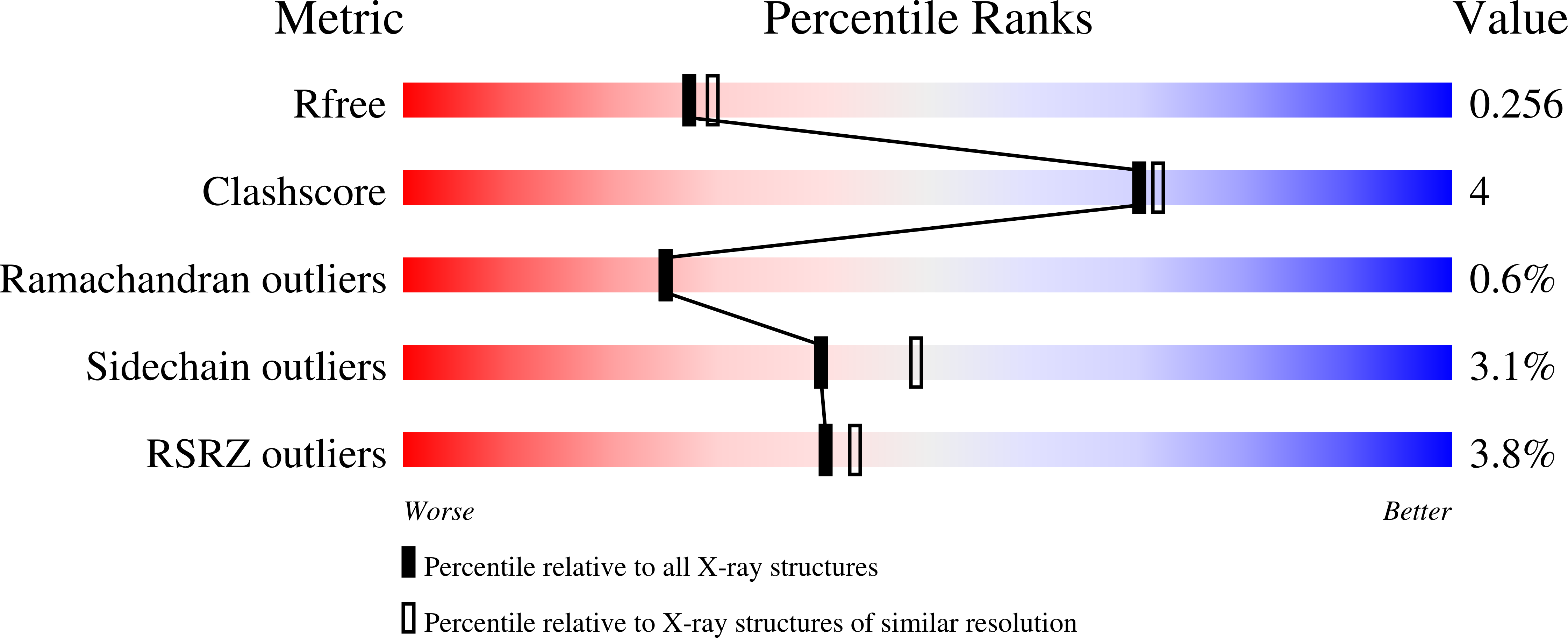
Resolution:
2.25 Å
R-Value Free:
0.25
R-Value Work:
0.19
R-Value Observed:
0.19
Space Group:
P 1
Macromolecular Entities
Structures with similar UniProt ID
Protein Blast
Polymer Type:polypeptide(L)
Molecule:Diphtheria toxin
Mutagens:K51E, E148K, H223Q, H257Q
Chain IDs:A, B
Chain Length:538
Number of Molecules:2
Biological Source:Corynebacterium diphtheriae
Primary Citation
Histidine Protonation and Conformational Switching in Diphtheria Toxin Translocation Domain.
Toxins 15 ? ? (2023)
PMID: 37505680 DOI: 10.3390/toxins15070410

Abstact

Protonation of key histidine residues has been long implicated in the acid-mediated cellular action of the diphtheria toxin translocation (T-) domain, responsible for the delivery of the catalytic domain into the cell. Here, we use a combination of computational (constant-pH Molecular Dynamics simulations) and experimental (NMR, circular dichroism, and fluorescence spectroscopy along with the X-ray crystallography) approaches to characterize the initial stages of conformational change happening in solution in the wild-type T-domain and in the H223Q/H257Q double mutant. This replacement suppresses the acid-induced transition, resulting in the retention of a more stable protein structure in solutions at pH 5.5 and, consequently, in reduced membrane-disrupting activity. Here, for the first time, we report the pKa values of the histidine residues of the T-domain, measured by NMR-monitored pH titrations. Most peaks in the histidine side chain spectral region are titrated with pKas ranging from 6.2 to 6.8. However, the two most up-field peaks display little change down to pH 6, which is a limiting pH for this protein in solution at concentrations required for NMR. These peaks are absent in the double mutant, suggesting they belong to H223 and H257. The constant-pH simulations indicate that for the T-domain in solution, the pKa values for histidine residues range from 3.0 to 6.5, with those most difficult to protonate being H251 and H257. Taken together, our experimental and computational data demonstrate that previously suggested cooperative protonation of all six histidines in the T-domain does not occur.

Legend

Protein

Chemical

Disease

Primary Citation of related structures