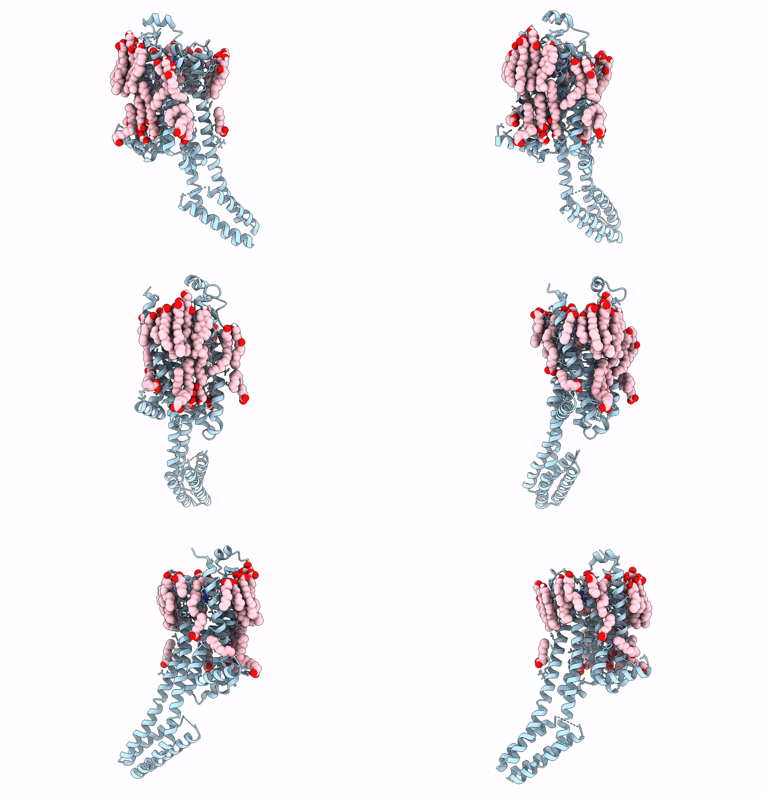
8FYN image
Deposition Date 2023-01-26
Release Date 2023-03-08
Last Version Date 2024-10-23
Entry Detail
PDB ID:
8FYN
Title:
MicroED structure of A2A from plasma milled lamellae
Biological Source:
Source Organism:
Homo sapiens (Taxon ID: 9606)
Method Details:
Experimental Method:
Resolution:
2.00 Å
R-Value Free:
0.27
R-Value Work:
0.22
R-Value Observed:
0.22
Space Group:
C 2 2 21
Macromolecular Entities
Polymer Type:polypeptide(L)
Molecule:Adenosine receptor A2a,Soluble cytochrome b562
Gene (Uniprot):cybC, ADORA2A
Chain IDs:A
Chain Length:447
Number of Molecules:1
Biological Source:Homo sapiens
Primary Citation
A robust approach for MicroED sample preparation of lipidic cubic phase embedded membrane protein crystals.
Nat Commun 14 1086 1086 (2023)
PMID: 36841804 DOI: 10.1038/s41467-023-36733-4

Abstact

Crystallizing G protein-coupled receptors (GPCRs) in lipidic cubic phase (LCP) often yields crystals suited for the cryogenic electron microscopy (cryoEM) method microcrystal electron diffraction (MicroED). However, sample preparation is challenging. Embedded crystals cannot be targeted topologically. Here, we use an integrated fluorescence light microscope (iFLM) inside of a focused ion beam and scanning electron microscope (FIB-SEM) to identify fluorescently labeled GPCR crystals. Crystals are targeted using the iFLM and LCP is milled using a plasma focused ion beam (pFIB). The optimal ion source for preparing biological lamellae is identified using standard crystals of proteinase K. Lamellae prepared using either argon or xenon produced the highest quality data and structures. MicroED data are collected from the milled lamellae and the structures are determined. This study outlines a robust approach to identify and mill membrane protein crystals for MicroED and demonstrates plasma ion-beam milling is a powerful tool for preparing biological lamellae.

Legend

Protein

Chemical

Disease

Primary Citation of related structures