Deposition Date
2023-01-25
Release Date
2023-08-30
Last Version Date
2024-10-16
Entry Detail
PDB ID:
8FXU
Keywords:
Title:
Thermoanaerobacter thermosaccharolyticum periplasmic Glucose-Binding Protein glucose complex: Badan conjugate attached at F17C
Biological Source:
Source Organism(s):
Thermoanaerobacterium thermosaccharolyticum (Taxon ID: 1517)
Expression System(s):
Method Details:
Experimental Method:
Resolution:
1.59 Å
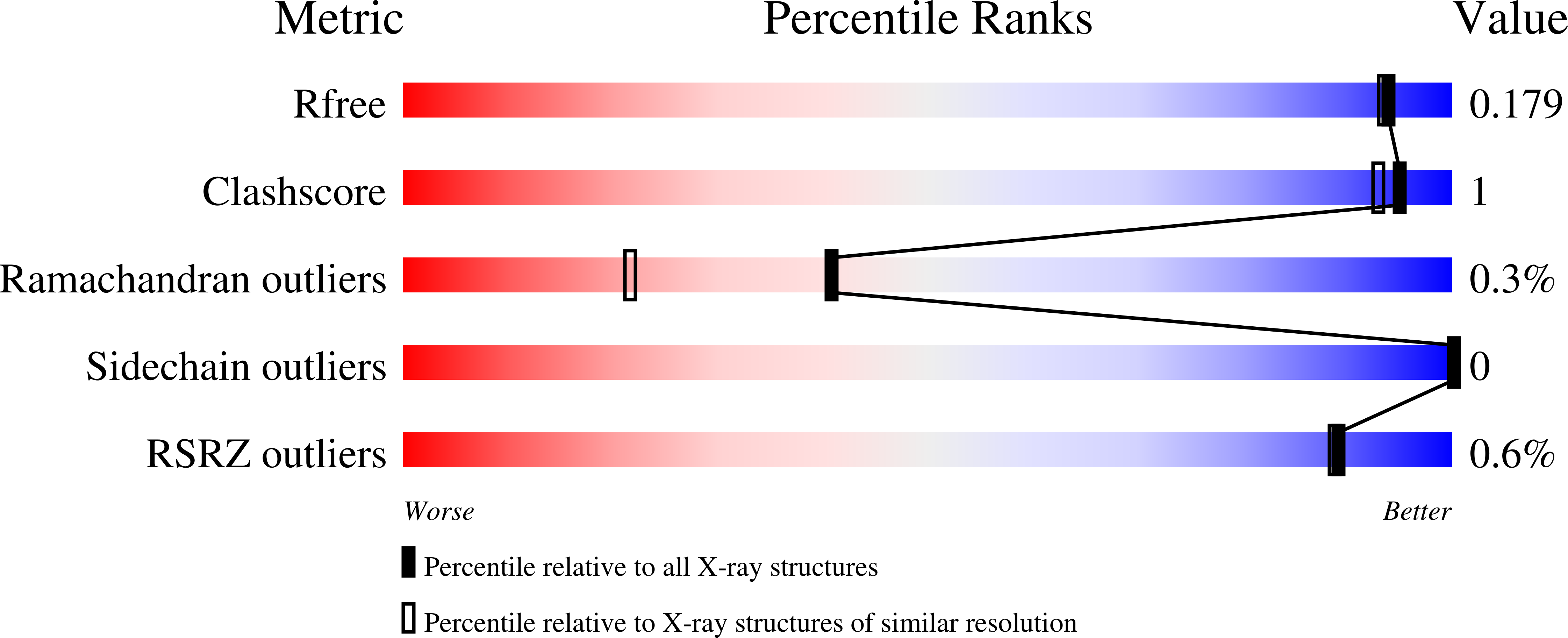
R-Value Free:
0.17
R-Value Work:
0.16
R-Value Observed:
0.16
Space Group:
P 21 21 21