
Deposition Date
2023-01-25
Release Date
2023-08-30
Last Version Date
2024-11-20
Entry Detail
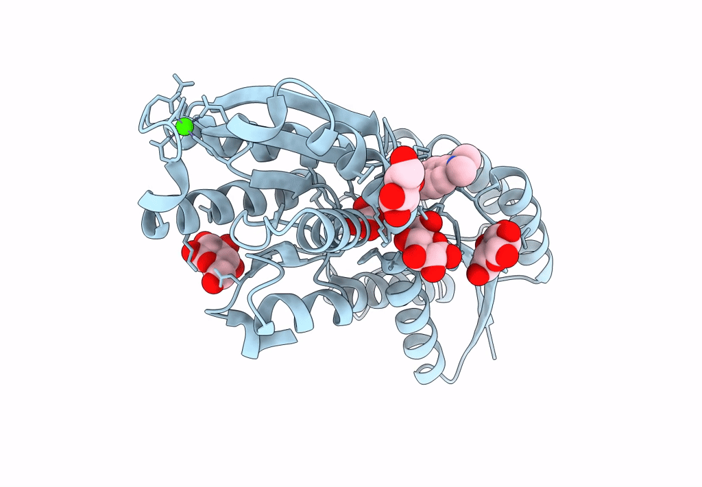
PDB ID:
8FXT
Keywords:
Title:
Escherichia coli periplasmic Glucose-Binding Protein glucose complex: Acrylodan conjugate attached at W183C
Biological Source:
Source Organism(s):
Escherichia coli (Taxon ID: 562)
Expression System(s):
Method Details:
Experimental Method:
Resolution:
1.53 Å
R-Value Free:
0.18
R-Value Work:
0.15
R-Value Observed:
0.15
Space Group:
C 1 2 1
