8FXF image
Entry Detail
PDB ID:
8FXF
Keywords:
Title:
Crystal structure of the coiled-coil domain of TRIM56
Biological Source:
Source Organism:
PDB Version:
Deposition Date:
2023-01-24
Release Date:
2023-05-24
Method Details:
Experimental Method:
Resolution:
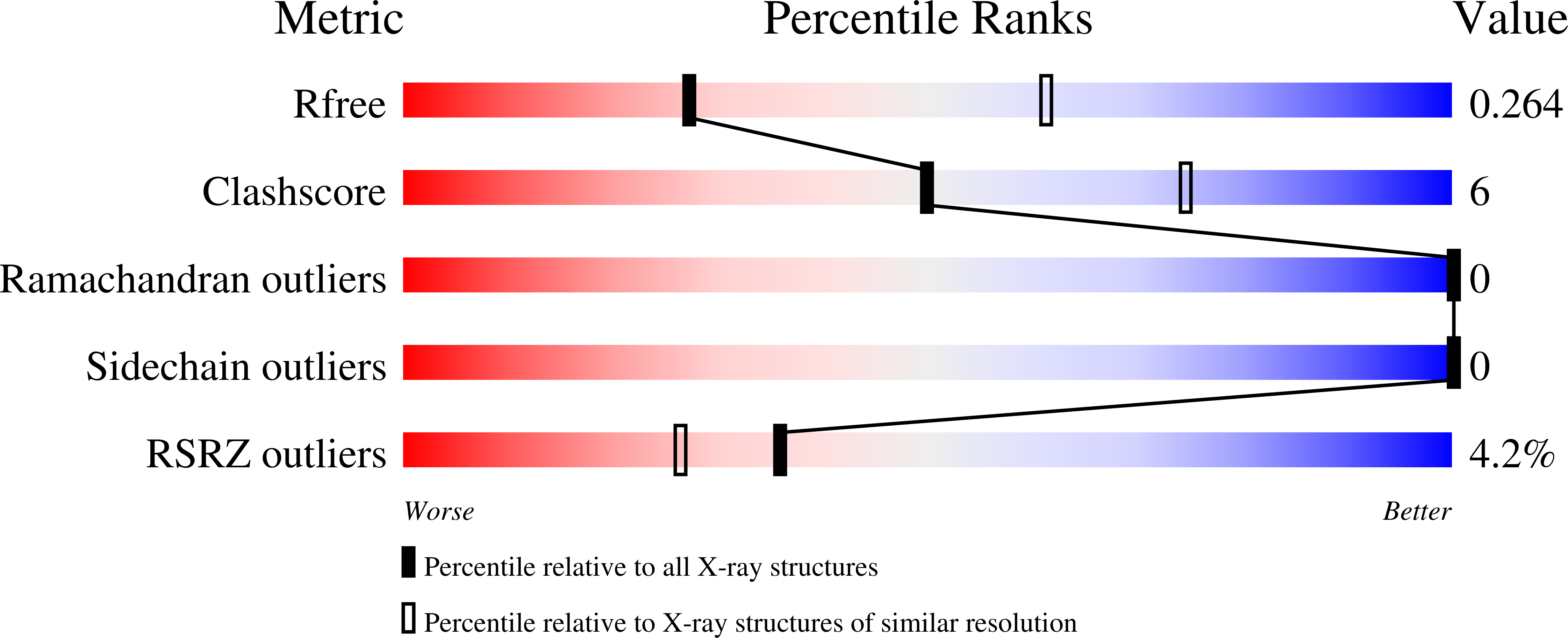
2.80 Å
R-Value Free:
0.26
R-Value Work:
0.21
Space Group:
I 41
Macromolecular Entities
Polymer Type:polypeptide(L)
Description:E3 ubiquitin-protein ligase TRIM56
Chain IDs:A, B, C, D
Chain Length:93
Number of Molecules:4
Biological Source:Mus musculus
Ligand Molecules
Primary Citation
TRIM56 coiled-coil domain structure provides insights into its E3 ligase functions.
Comput Struct Biotechnol J 21 2801 2808 (2023)
PMID: 37168870 DOI: 10.1016/j.csbj.2023.04.022

Abstact

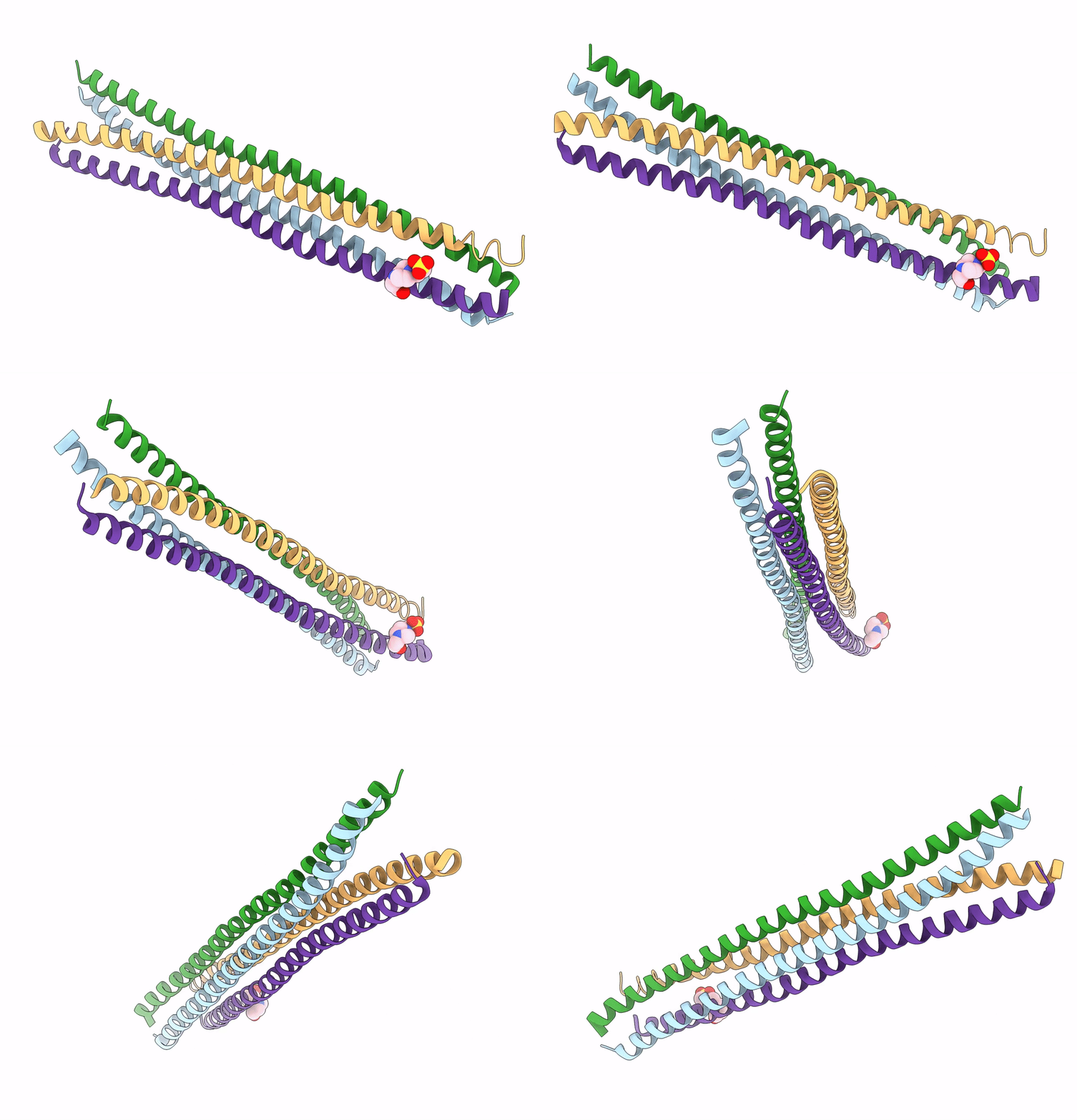
Protein ubiquitination is a post-translation modification mediated by E3 ubiquitin ligases. The RING domain E3 ligases are the largest family of E3 ubiquitin ligases, they act as a scaffold, bringing the E2-ubiquitin complex and its substrate together to facilitate direct ubiquitin transfer. However, the quaternary structures of RING E3 ligases that perform ubiquitin transfer remain poorly understood. In this study, we solved the crystal structure of TRIM56, a member of the RING E3 ligase. The structure of the coiled-coil domain indicated that the two anti-parallel dimers bound together to form a tetramer at a small crossing angle. This tetramer structure allows two RING domains to exist on each side to form an active homodimer in supporting ubiquitin transfer from E2 to its nearby substrate recruited by the C-terminal domains on the same side. These findings suggest that the coiled-coil domain-mediated tetramer is a feasible scaffold for facilitating the recruitment and transfer of ubiquitin to accomplish E3 ligase activity.

Legend

Protein

Chemical

Disease

Primary Citation of related structures