8FWL image
Deposition Date 2023-01-23
Release Date 2023-03-08
Last Version Date 2024-03-20
Entry Detail
PDB ID:
8FWL
Keywords:
Title:
Crystal structure of Australian bat lyssavirus nucleoprotein in complex with phosphoprotein chaperone
Biological Source:
Source Organism:
Method Details:
Experimental Method:
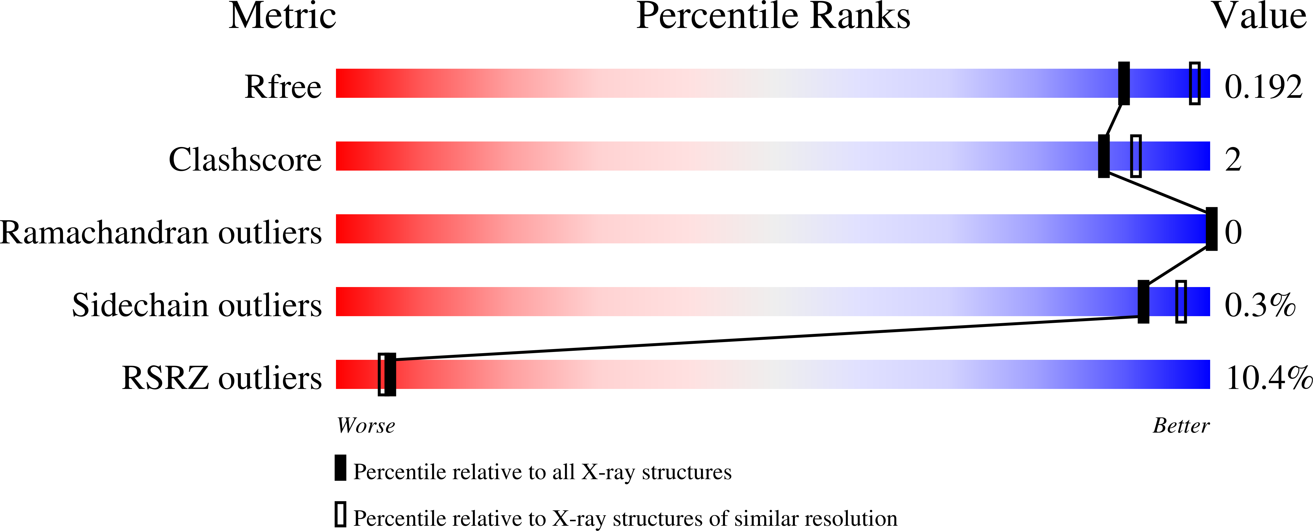
Resolution:
2.19 Å
R-Value Free:
0.19
R-Value Work:
0.15
R-Value Observed:
0.16
Space Group:
P 1 2 1
Macromolecular Entities
Structures with similar UniProt ID
Protein Blast
Polymer Type:polypeptide(L)
Molecule:Phosphoprotein,Nucleoprotein
Chain IDs:A
Chain Length:567
Number of Molecules:1
Biological Source:Lyssavirus australis
Ligand Molecules
Primary Citation
Structural Determination of the Australian Bat Lyssavirus Nucleoprotein and Phosphoprotein Complex.
Viruses 16 33 33 (2023)
PMID: 38229694 DOI: 10.3390/v16010033

Abstact

Australian bat lyssavirus (ABLV) shows similar clinical symptoms as rabies, but there are currently no protein structures available for ABLV proteins. In lyssaviruses, the interaction between nucleoprotein (N) and phosphoprotein (N) in the absence of RNA generates a complex (N0P) that is crucial for viral assembly, and understanding the interface between these two proteins has the potential to provide insight into a key feature: the viral lifecycle. In this study, we used recombinant chimeric protein expression and X-ray crystallography to determine the structure of ABLV nucleoprotein bound to residues 1-40 of its phosphoprotein chaperone. Comparison of our results with the recently generated structure of RABV CVS-11 N0P demonstrated a highly conserved interface in this complex. Because the N0P interface is conserved in the lyssaviruses of phylogroup I, it is an attractive therapeutic target for multiple rabies-causing viral species.

Legend

Protein

Chemical

Disease

Primary Citation of related structures
Feedback Form
Name
Email
Institute
Feedback