8FTY image
Entry Detail
PDB ID:
8FTY
Keywords:
Title:
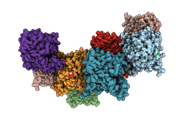
Crystal structure of the carotenoid isomerooxygenase, NinaB
Biological Source:
Source Organism:
Host Organism:
PDB Version:
Deposition Date:
2023-01-14
Release Date:
2024-01-17
Method Details:
Experimental Method:
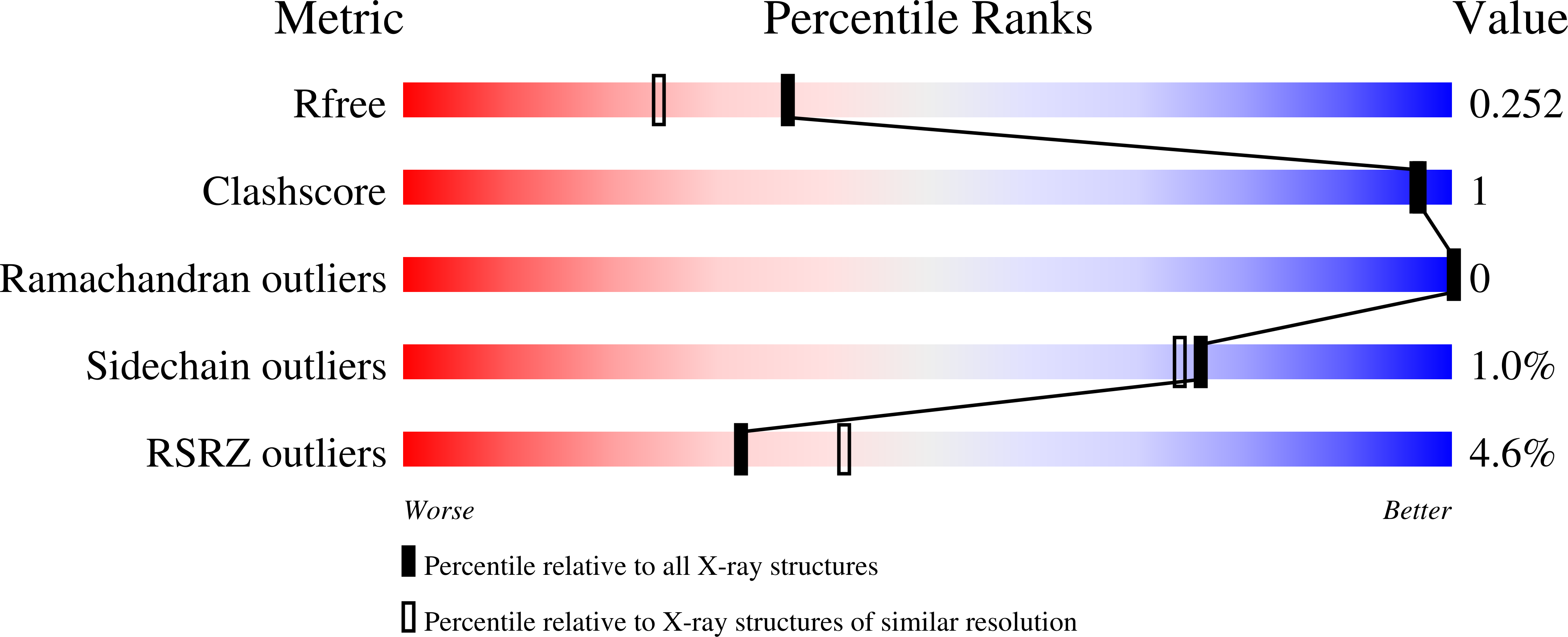
Resolution:
1.95 Å
R-Value Free:
0.24
R-Value Work:
0.21
R-Value Observed:
0.21
Space Group:
C 1 2 1
Macromolecular Entities
Polymer Type:polypeptide(L)
Description:carotenoid isomerooxygenase
Chain IDs:A, B, C, D
Chain Length:509
Number of Molecules:4
Biological Source:Trichoplusia ni
Polymer Type:polypeptide(L)
Description:carotenoid isomerooxygenase
Chain IDs:E, F
Chain Length:509
Number of Molecules:2
Biological Source:Trichoplusia ni
Polymer Type:polypeptide(L)
Description:carotenoid isomerooxygenase
Chain IDs:G, H
Chain Length:509
Number of Molecules:2
Biological Source:Trichoplusia ni
Modified Residue
Compound ID Chain ID Parent Comp ID Details 2D Image
CSD G CYS modified residue
CSO E CYS modified residue
Primary Citation
Carotenoid cleavage enzymes evolved convergently to generate the visual chromophore.
Nat.Chem.Biol. 20 779 788 (2024)
PMID: 38355721 DOI: 10.1038/s41589-024-01554-z

Abstact

The retinal light response in animals originates from the photoisomerization of an opsin-coupled 11-cis-retinaldehyde chromophore. This visual chromophore is enzymatically produced through the action of carotenoid cleavage dioxygenases. Vertebrates require two carotenoid cleavage dioxygenases, β-carotene oxygenase 1 and retinal pigment epithelium 65 (RPE65), to form 11-cis-retinaldehyde from carotenoid substrates, whereas invertebrates such as insects use a single enzyme known as Neither Inactivation Nor Afterpotential B (NinaB). RPE65 and NinaB couple trans-cis isomerization with hydrolysis and oxygenation, respectively, but the mechanistic relationship of their isomerase activities remains unknown. Here we report the structure of NinaB, revealing details of its active site architecture and mode of membrane binding. Structure-guided mutagenesis studies identify a residue cluster deep within the NinaB substrate-binding cleft that controls its isomerization activity. Our data demonstrate that isomerization activity is mediated by distinct active site regions in NinaB and RPE65-an evolutionary convergence that deepens our understanding of visual system diversity.

Legend

Protein

Chemical

Disease

Primary Citation of related structures