8FO8 image
Deposition Date 2022-12-29
Release Date 2024-01-03
Last Version Date 2024-01-03
Entry Detail
PDB ID:
8FO8
Keywords:
Title:
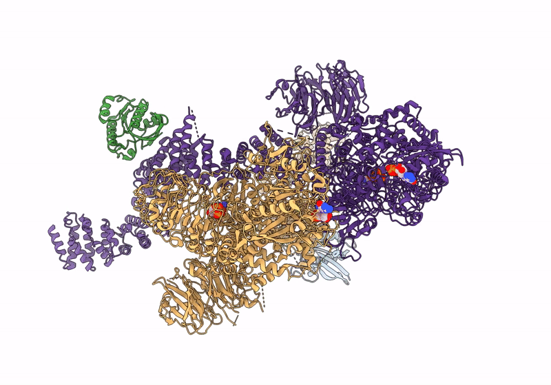
Cryo-EM structure of Rab29-LRRK2 complex in the LRRK2 dimer state
Biological Source:
Source Organism:
Homo sapiens (Taxon ID: 9606)
Host Organism:
Method Details:
Experimental Method:
Resolution:
3.88 Å
Aggregation State:
PARTICLE
Reconstruction Method:
SINGLE PARTICLE
Macromolecular Entities
Polymer Type:polypeptide(L)
Molecule:Ras-related protein Rab-7L1
Gene (Uniprot):RAB29
Chain IDs:A, B
Chain Length:177
Number of Molecules:2
Biological Source:Homo sapiens
Polymer Type:polypeptide(L)
Molecule:Leucine-rich repeat serine/threonine-protein kinase 2
Gene (Uniprot):LRRK2
Chain IDs:C, D (auth: E)
Chain Length:2527
Number of Molecules:2
Biological Source:Homo sapiens
Primary Citation
Rab29-dependent asymmetrical activation of leucine-rich repeat kinase 2.
Science 382 1404 1411 (2023)
PMID: 38127736 DOI: 10.1126/science.adi9926

Abstact

Gain-of-function mutations in LRRK2, which encodes the leucine-rich repeat kinase 2 (LRRK2), are the most common genetic cause of late-onset Parkinson's disease. LRRK2 is recruited to membrane organelles and activated by Rab29, a Rab guanosine triphosphatase encoded in the PARK16 locus. We present cryo-electron microscopy structures of Rab29-LRRK2 complexes in three oligomeric states, providing key snapshots during LRRK2 recruitment and activation. Rab29 induces an unexpected tetrameric assembly of LRRK2, formed by two kinase-active central protomers and two kinase-inactive peripheral protomers. The central protomers resemble the active-like state trapped by the type I kinase inhibitor DNL201, a compound that underwent a phase 1 clinical trial. Our work reveals the structural mechanism of LRRK2 spatial regulation and provides insights into LRRK2 inhibitor design for Parkinson's disease treatment.

Legend

Protein

Chemical

Disease

Primary Citation of related structures