Deposition Date
2022-12-21
Release Date
2024-01-10
Last Version Date
2024-10-09
Entry Detail
PDB ID:
8FL5
Keywords:
Title:
Crystal Structure of Enterovirus 68 3C Protease inactive mutant C147A at 1.8 Angstroms.
Biological Source:
Source Organism(s):
enterovirus D68 (Taxon ID: 42789)
Expression System(s):
Method Details:
Experimental Method:
Resolution:
1.80 Å
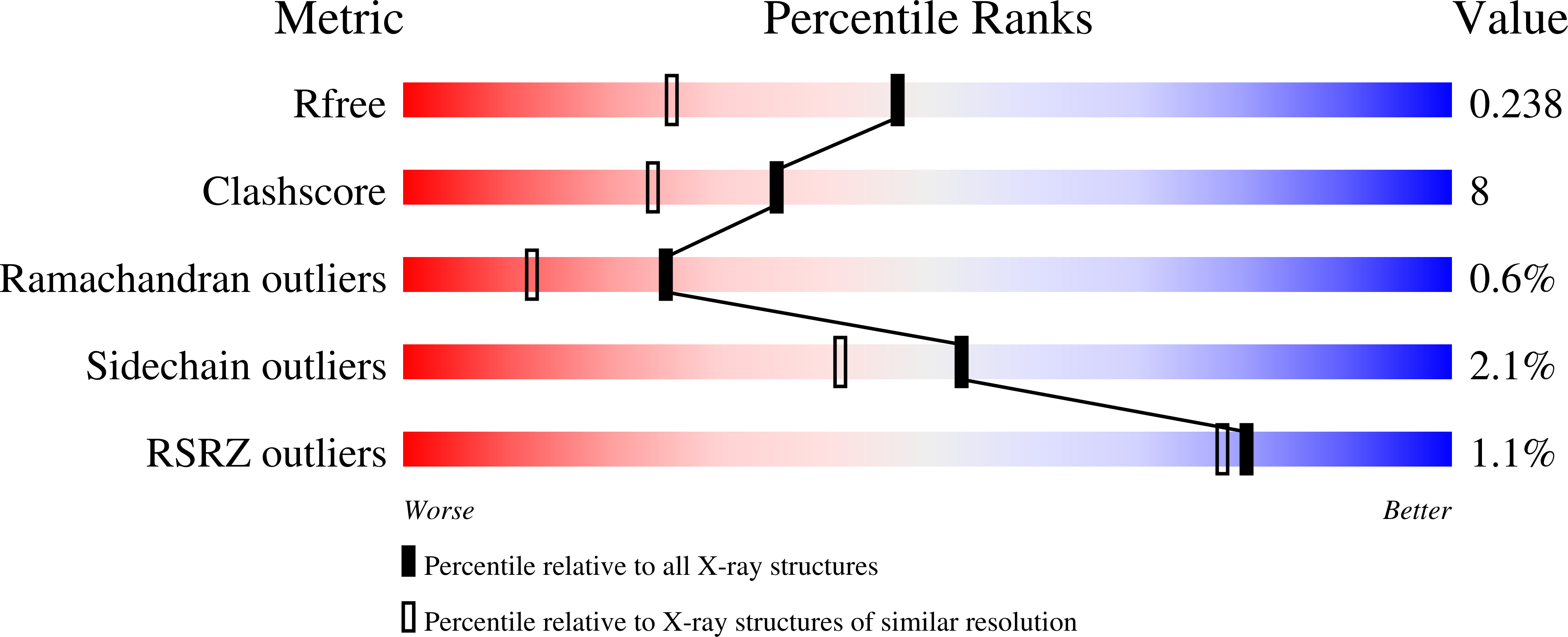
R-Value Free:
0.23
R-Value Work:
0.17
R-Value Observed:
0.17
Space Group:
P 1 21 1