
Deposition Date
2022-11-22
Release Date
2023-06-07
Last Version Date
2023-07-05
Entry Detail
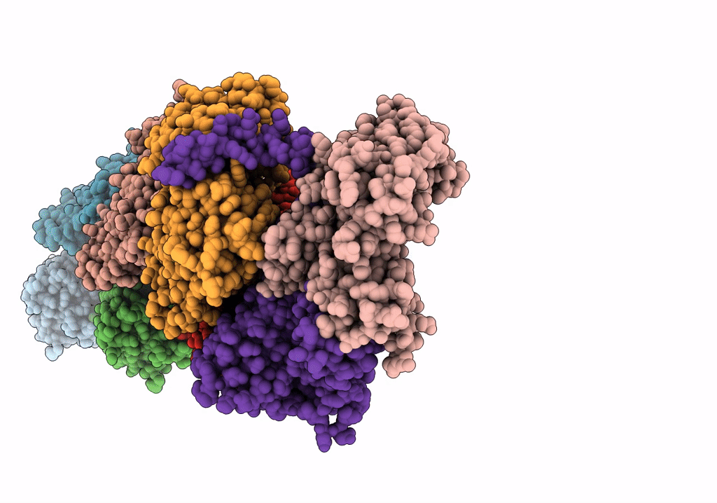
PDB ID:
8F8Q
Keywords:
Title:
Cryo-EM structure of the CapZ-capped barbed end of F-actin
Biological Source:
Source Organism(s):
Homo sapiens (Taxon ID: 9606)
Oryctolagus cuniculus (Taxon ID: 9986)
Oryctolagus cuniculus (Taxon ID: 9986)
Expression System(s):
Method Details:
Experimental Method:
Resolution:
2.79 Å
Aggregation State:
PARTICLE
Reconstruction Method:
SINGLE PARTICLE
