8F8N image
Deposition Date 2022-11-22
Release Date 2023-08-16
Last Version Date 2024-11-06
Entry Detail
PDB ID:
8F8N
Title:
Crystal structure of the Arabidopsis SPIRAL2 C-terminal domain
Biological Source:
Source Organism:
Method Details:
Experimental Method:
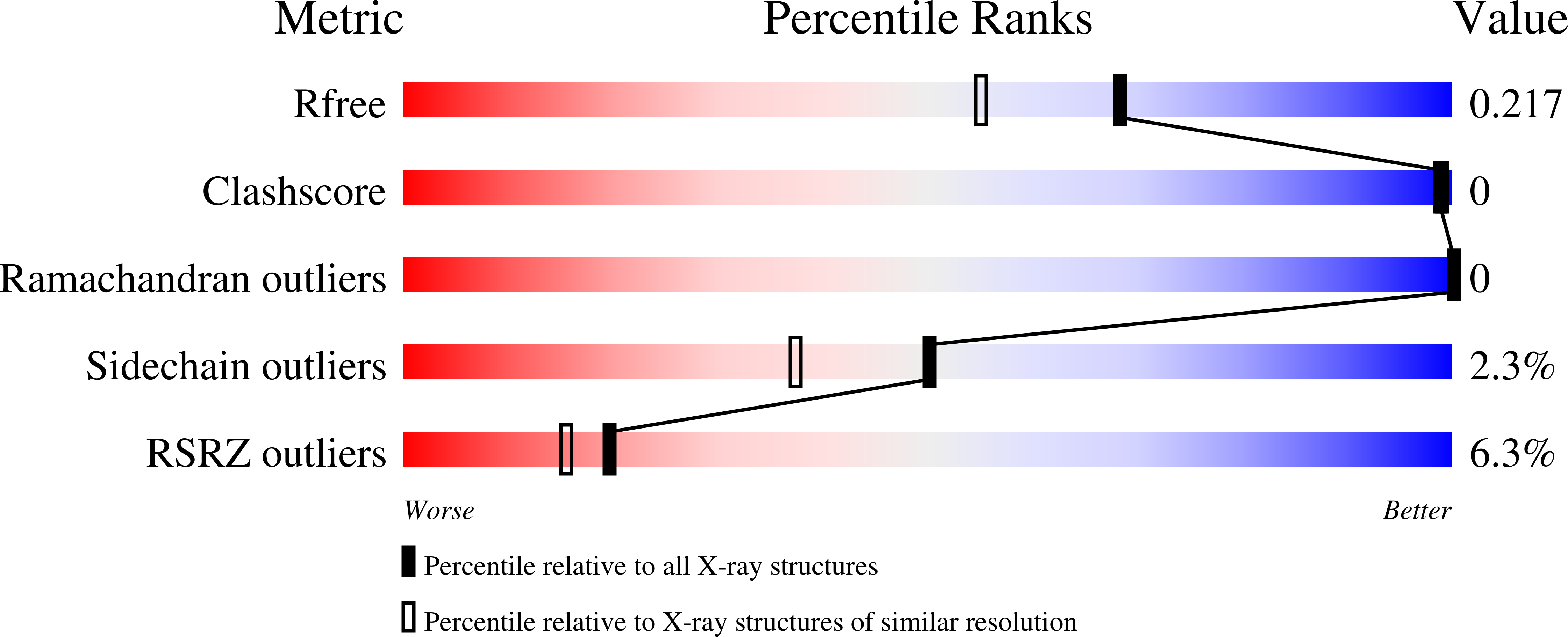
Resolution:
1.80 Å
R-Value Free:
0.21
R-Value Work:
0.18
R-Value Observed:
0.18
Space Group:
P 21 21 21
Macromolecular Entities
Structures with similar UniProt ID
Protein Blast
Polymer Type:polypeptide(L)
Molecule:Microtubule-associated protein TORTIFOLIA1
Gene (Uniprot):TOR1
Chain IDs:A
Chain Length:220
Number of Molecules:1
Biological Source:Arabidopsis thaliana
Modified Residue
Compound ID Chain ID Parent Comp ID Details 2D Image
MSE A MET modified residue
Primary Citation
Crystal structure of the Arabidopsis SPIRAL2 C-terminal domain reveals a p80-Katanin-like domain.
Plos One 18 e0290024 e0290024 (2023)
PMID: 38157339 DOI: 10.1371/journal.pone.0290024

Abstact

Epidermal cells of dark-grown plant seedlings reorient their cortical microtubule arrays in response to blue light from a net lateral orientation to a net longitudinal orientation with respect to the long axis of cells. The molecular mechanism underlying this microtubule array reorientation involves katanin, a microtubule severing enzyme, and a plant-specific microtubule associated protein called SPIRAL2. Katanin preferentially severs longitudinal microtubules, generating seeds that amplify the longitudinal array. Upon severing, SPIRAL2 binds nascent microtubule minus ends and limits their dynamics, thereby stabilizing the longitudinal array while the lateral array undergoes net depolymerization. To date, no experimental structural information is available for SPIRAL2 to help inform its mechanism. To gain insight into SPIRAL2 structure and function, we determined a 1.8 Å resolution crystal structure of the Arabidopsis thaliana SPIRAL2 C-terminal domain. The domain is composed of seven core α-helices, arranged in an α-solenoid. Amino-acid sequence conservation maps primarily to one face of the domain involving helices α1, α3, α5, and an extended loop, the α6-α7 loop. The domain fold is similar to, yet structurally distinct from the C-terminal domain of Ge-1 (an mRNA decapping complex factor involved in P-body localization) and, surprisingly, the C-terminal domain of the katanin p80 regulatory subunit. The katanin p80 C-terminal domain heterodimerizes with the MIT domain of the katanin p60 catalytic subunit, and in metazoans, binds the microtubule minus-end factors CAMSAP3 and ASPM. Structural analysis predicts that SPIRAL2 does not engage katanin p60 in a mode homologous to katanin p80. The SPIRAL2 structure highlights an interesting evolutionary convergence of domain architecture and microtubule minus-end localization between SPIRAL2 and katanin complexes, and establishes a foundation upon which structure-function analysis can be conducted to elucidate the role of this domain in the regulation of plant microtubule arrays.

Legend

Protein

Chemical

Disease

Primary Citation of related structures