
Deposition Date
2022-10-26
Release Date
2023-07-05
Last Version Date
2024-10-23
Entry Detail
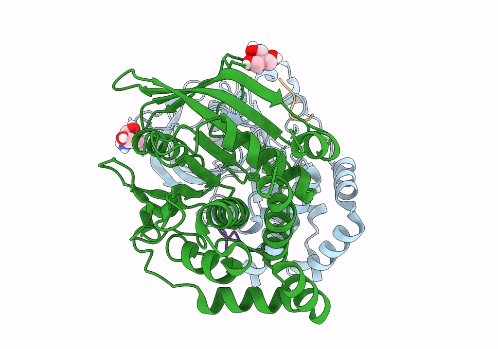
PDB ID:
8EYB
Keywords:
Title:
Crystal structure of PTP1B D181A/Q262A/C215A phosphatase domain with JAK2 activation loop phosphopeptide
Biological Source:
Source Organism(s):
Homo sapiens (Taxon ID: 9606)
Expression System(s):
Method Details:
Experimental Method:
Resolution:
2.35 Å
R-Value Free:
0.24
R-Value Work:
0.20
R-Value Observed:
0.20
Space Group:
P 31 2 1
