Deposition Date
2022-10-20
Release Date
2023-03-01
Last Version Date
2024-05-01
Entry Detail
PDB ID:
8EVK
Keywords:
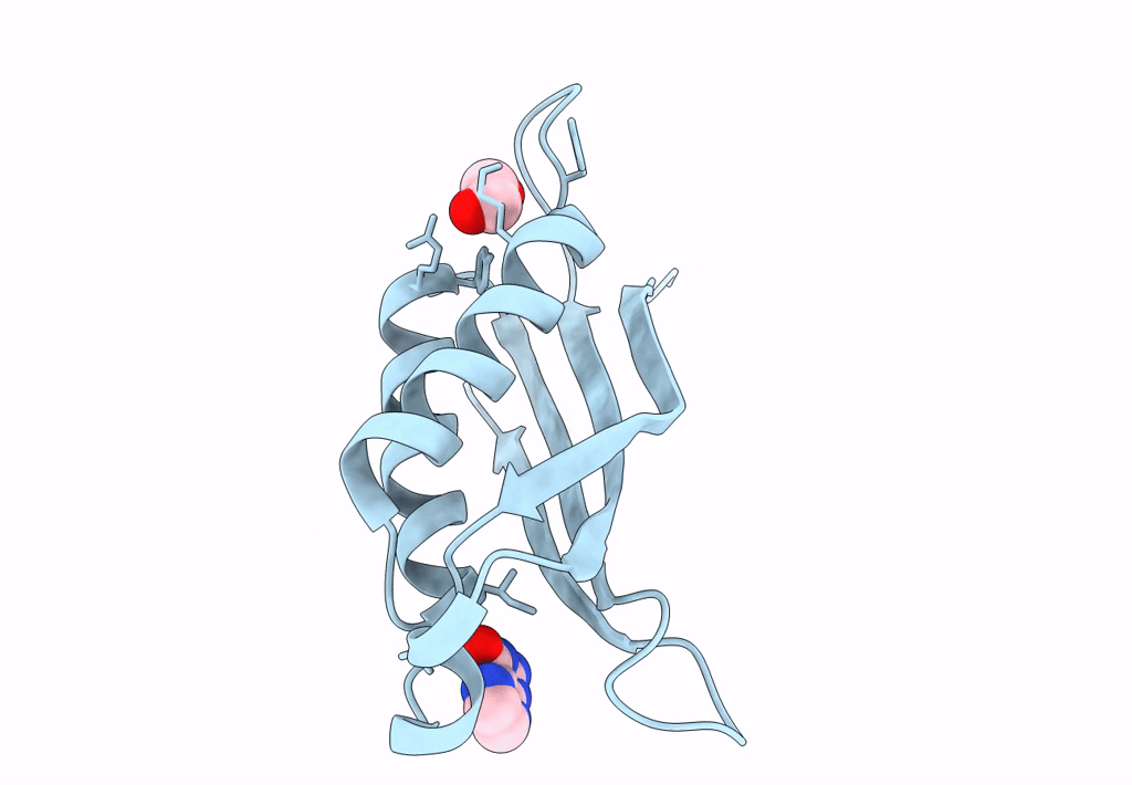
Title:
Crystal structure of Helicobacter pylori dihydroneopterin aldolase (DHNA)
Biological Source:
Source Organism(s):
Helicobacter pylori G27 (Taxon ID: 563041)
Expression System(s):
Method Details:
Experimental Method:
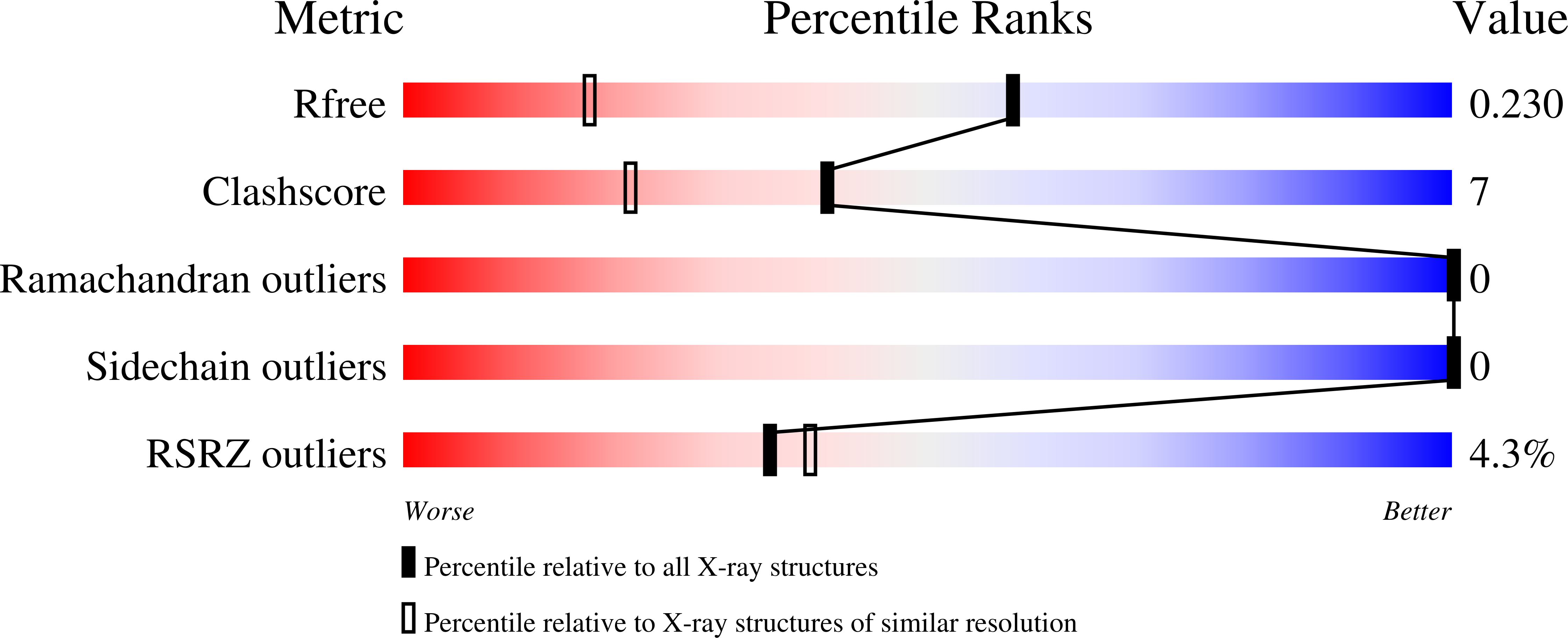
Resolution:
1.49 Å
R-Value Free:
0.23
R-Value Work:
0.19
R-Value Observed:
0.19
Space Group:
I 4