8EQ8 image
Entry Detail
PDB ID:
8EQ8
Keywords:
Title:
The crystal structure of 14-3-3 Beta containing 3-nitrotyrosine at position Y130
Biological Source:
Source Organism:
Host Organism:
PDB Version:
Deposition Date:
2022-10-07
Release Date:
2023-01-25
Method Details:
Experimental Method:
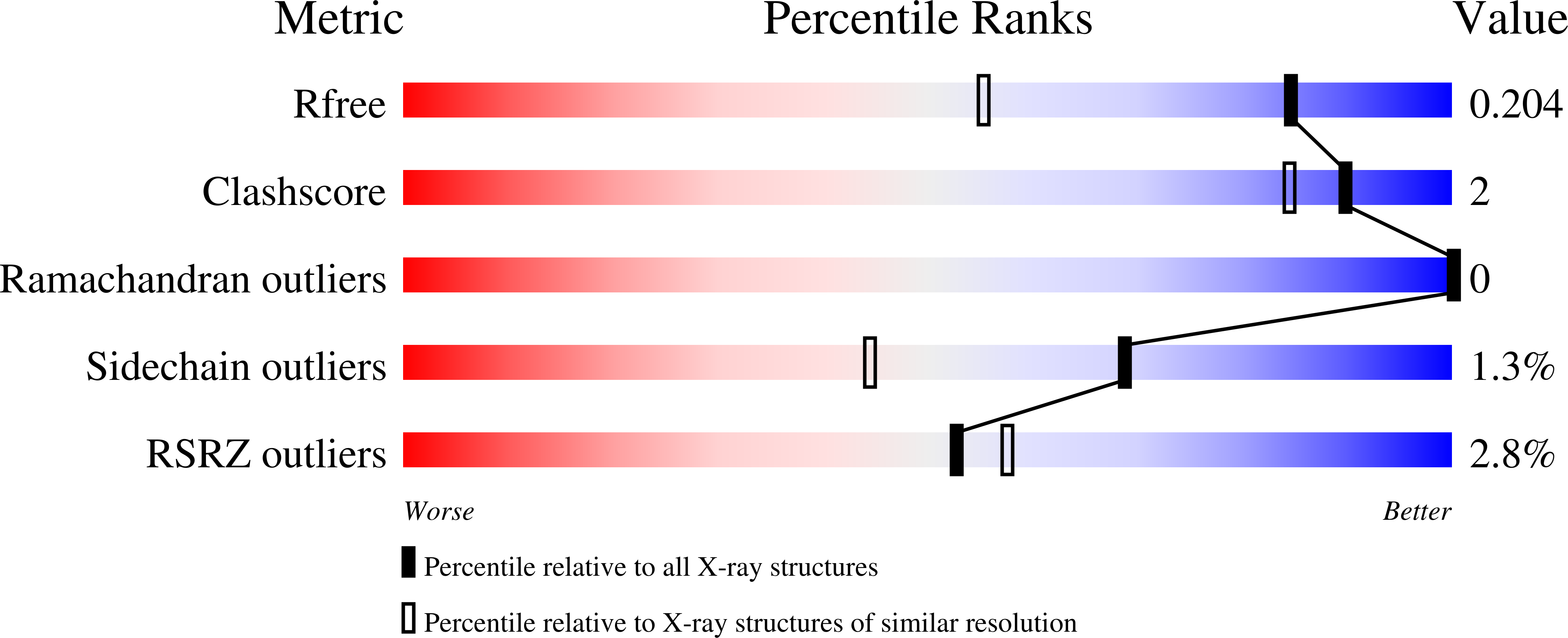
Resolution:
1.50 Å
R-Value Free:
0.20
R-Value Work:
0.17
R-Value Observed:
0.17
Space Group:
P 1 21 1
Macromolecular Entities
Polymer Type:polypeptide(L)
Description:14-3-3 protein beta/alpha
Chain IDs:A (auth: B), B (auth: A)
Chain Length:245
Number of Molecules:2
Biological Source:Homo sapiens
Modified Residue
Compound ID Chain ID Parent Comp ID Details 2D Image
NIY A TYR modified residue
Primary Citation
Genetic encoding of 3-nitro-tyrosine reveals the impacts of 14-3-3 nitration on client binding and dephosphorylation.
Protein Sci. 32 e4574 e4574 (2023)
PMID: 36691781 DOI: 10.1002/pro.4574

Abstact

14-3-3 proteins are central hub regulators of hundreds of phosphorylated "client" proteins. They are subject to over 60 post-translational modifications (PTMs), yet little is known how these PTMs alter 14-3-3 function and its ability to regulate downstream signaling pathways. An often neglected, but well-documented 14-3-3 PTM found under physiological and immune-stimulatory conditions is the conversion of tyrosine to 3-nitro-tyrosine at several Tyr sites, two of which are located at sites considered important for 14-3-3 function: Y130 (β-isoform numbering) is located in the primary phospho-client peptide-binding groove, while Y213 is found on a secondary binding site that engages with clients for full 14-3-3/client complex formation and client regulation. By genetically encoding 3-nitro-tyrosine, we sought to understand if nitration at Y130 and Y213 effectively modulated 14-3-3 structure, function, and client complexation. The 1.5 Å resolution crystal structure of 14-3-3 nitrated at Y130 showed the nitro group altered the conformation of key residues in the primary binding site, while functional studies confirmed client proteins failed to bind this variant of 14-3-3. But, in contrast to other client-binding deficient variants, it did not localize to the nucleus. The 1.9 Å resolution structure of 14-3-3 nitrated at Y213 revealed unusual flexibility of its C-terminal α-helix resulting in domain swapping, suggesting additional structural plasticity though its relevance is not clear as this nitrated form retained its ability to bind clients. Collectively, our data suggest that nitration of 14-3-3 will alter downstream signaling systems, and if uncontrolled could result in global dysregulation of the 14-3-3 interactome.

Legend

Protein

Chemical

Disease

Primary Citation of related structures