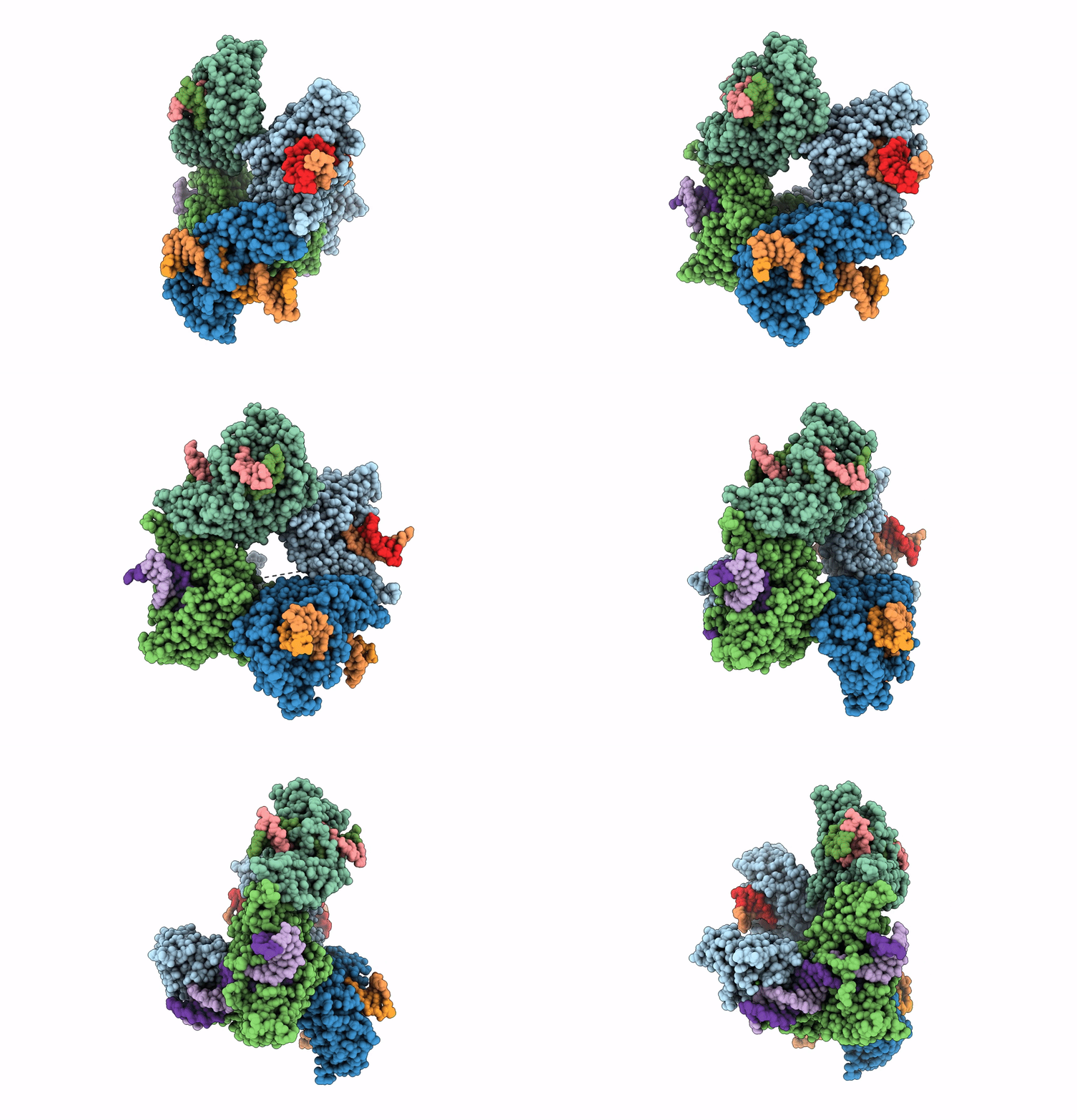
8EPX image
Entry Detail
PDB ID:
8EPX
EMDB ID:
Title:
Type IIS Restriction Endonuclease PaqCI, DNA bound
Biological Source:
Host Organism:
PDB Version:
Deposition Date:
2022-10-06
Release Date:
2023-03-22
Method Details:
Experimental Method:
Resolution:
3.15 Å
Aggregation State:
PARTICLE
Reconstruction Method:
SINGLE PARTICLE
Macromolecular Entities
Polymer Type:polypeptide(L)
Description:Type IIS Restriction Endonuclease PaqCI
Chain IDs:A, B, C, D
Chain Length:510
Number of Molecules:4
Biological Source:Paucibacter aquatile
Polymer Type:polydeoxyribonucleotide
Description:DNA 4a
Chain IDs:E
Chain Length:15
Number of Molecules:1
Biological Source:DNA molecule
Polymer Type:polydeoxyribonucleotide
Description:DNA 4b
Chain IDs:F
Chain Length:15
Number of Molecules:1
Biological Source:DNA molecule
Polymer Type:polydeoxyribonucleotide
Description:DNA 2b
Chain IDs:H (auth: G)
Chain Length:16
Number of Molecules:1
Biological Source:DNA molecule
Polymer Type:polydeoxyribonucleotide
Description:DNA 2a
Chain IDs:G (auth: H)
Chain Length:16
Number of Molecules:1
Biological Source:DNA molecule
Polymer Type:polydeoxyribonucleotide
Description:DNA 3a
Chain IDs:I
Chain Length:18
Number of Molecules:1
Biological Source:DNA molecule
Polymer Type:polydeoxyribonucleotide
Description:DNA 3b
Chain IDs:J
Chain Length:18
Number of Molecules:1
Biological Source:DNA molecule
Polymer Type:polydeoxyribonucleotide
Description:DNA 1a
Chain IDs:K
Chain Length:23
Number of Molecules:1
Biological Source:DNA molecule
Polymer Type:polydeoxyribonucleotide
Description:DNA 1b
Chain IDs:L
Chain Length:23
Number of Molecules:1
Biological Source:DNA molecule
Ligand Molecules
Primary Citation
Structures, activity and mechanism of the Type IIS restriction endonuclease PaqCI.
Nucleic Acids Res. 51 4467 4487 (2023)
PMID: 36987874 DOI: 10.1093/nar/gkad228

Abstact

Type IIS restriction endonucleases contain separate DNA recognition and catalytic domains and cleave their substrates at well-defined distances outside their target sequences. They are employed in biotechnology for a variety of purposes, including the creation of gene-targeting zinc finger and TAL effector nucleases and DNA synthesis applications such as Golden Gate assembly. The most thoroughly studied Type IIS enzyme, FokI, has been shown to require multimerization and engagement with multiple DNA targets for optimal cleavage activity; however, details of how it or similar enzymes forms a DNA-bound reaction complex have not been described at atomic resolution. Here we describe biochemical analyses of DNA cleavage by the Type IIS PaqCI restriction endonuclease and a series of molecular structures in the presence and absence of multiple bound DNA targets. The enzyme displays a similar tetrameric organization of target recognition domains in the absence or presence of bound substrate, with a significant repositioning of endonuclease domains in a trapped DNA-bound complex that is poised to deliver the first of a series of double-strand breaks. PaqCI and FokI share similar structural mechanisms of DNA cleavage, but considerable differences in their domain organization and quaternary architecture, facilitating comparisons between distinct Type IIS enzymes.

Legend

Protein

Chemical

Disease

Primary Citation of related structures