8ENC image
Deposition Date 2022-09-29
Release Date 2022-11-23
Last Version Date 2024-06-19
Entry Detail
PDB ID:
8ENC
Keywords:
Title:
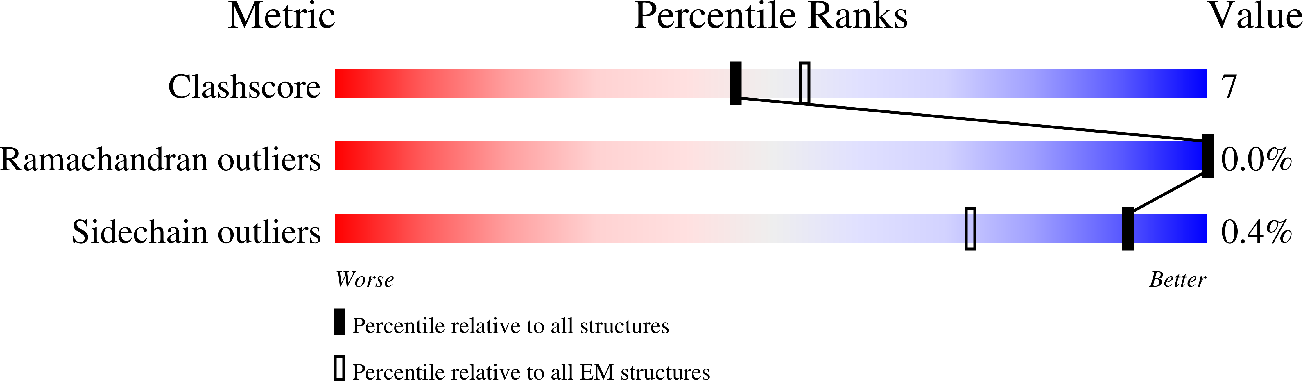
Helical reconstruction of the human cardiac actin-tropomyosin-myosin loop 4 7G mutant complex
Biological Source:
Source Organism:
Homo sapiens (Taxon ID: 9606)
Sus scrofa (Taxon ID: 9823)
Host Organism:
Method Details:
Experimental Method:
Resolution:
3.60 Å
Aggregation State:
FILAMENT
Reconstruction Method:
HELICAL
Macromolecular Entities
Polymer Type:polypeptide(L)
Molecule:Actin, alpha cardiac muscle 1
Gene (Uniprot):ACTC1
Chain IDs:B, C, D, E, F
Chain Length:377
Number of Molecules:5
Biological Source:Sus scrofa
Polymer Type:polypeptide(L)
Molecule:Myosin-7
Gene (Uniprot):MYH7
Mutations:Residues 366-372 substituted with seven glycines
Chain IDs:A (auth: M)
Chain Length:1935
Number of Molecules:1
Biological Source:Homo sapiens
Polymer Type:polypeptide(L)
Molecule:Tropomyosin alpha-1 chain
Gene (Uniprot):TPM1
Chain IDs:G (auth: O), H (auth: P)
Chain Length:284
Number of Molecules:2
Biological Source:Homo sapiens
Primary Citation
Myosin loop-4 is critical for optimal tropomyosin repositioning on actin during muscle activation and relaxation.
J.Gen.Physiol. 155 ? ? (2023)
PMID: 36459134 DOI: 10.1085/jgp.202213274

Abstact

During force-generating steps of the muscle crossbridge cycle, the tip of the myosin motor, specifically loop-4, contacts the tropomyosin cable of actin filaments. In the current study, we determined the corresponding effect of myosin loop-4 on the regulatory positioning of tropomyosin on actin. To accomplish this, we compared high-resolution cryo-EM structures of myosin S1-decorated thin filaments containing either wild-type or a loop-4 mutant construct, where the seven-residue portion of myosin loop-4 that contacts tropomyosin was replaced by glycine residues, thus removing polar side chains from residues 366-372. Cryo-EM analysis of fully decorated actin-tropomyosin filaments with wild-type and mutant S1, yielded 3.4-3.6 Å resolution reconstructions, with even higher definition at the actin-myosin interface. Loop-4 densities both in wild-type and mutant S1 were clearly identified, and side chains were resolved in the wild-type structure. Aside from loop-4, actin and myosin structural domains were indistinguishable from each other when filaments were decorated with either mutant or wild-type S1. In marked contrast, the position of tropomyosin on actin in the two reconstructions differed by 3 to 4 Å. In maps of filaments containing the mutant, tropomyosin was located closer to the myosin-head and thus moved in the direction of the C-state conformation adopted by myosin-free thin filaments. Complementary interaction energy measurements showed that tropomyosin in the mutant thin filaments sits on actin in a local energy minimum, whereas tropomyosin is positioned by wild-type S1 in an energetically unfavorable location. We propose that the high potential energy associated with tropomyosin positioning in wild-type filaments favors an effective transition to B- and C-states following release of myosin from the thin filaments during relaxation.

Legend

Protein

Chemical

Disease

Primary Citation of related structures