Deposition Date
2022-09-28
Release Date
2023-05-24
Last Version Date
2024-09-25
Entry Detail
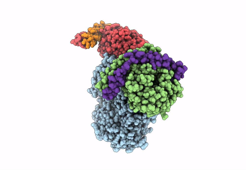
PDB ID:
8EMW
Keywords:
Title:
Phospholipase C beta 3 (PLCb3) in complex with Gbg on liposomes
Biological Source:
Source Organism(s):
Homo sapiens (Taxon ID: 9606)
Expression System(s):
Method Details:
Experimental Method:
Resolution:
3.50 Å
Aggregation State:
PARTICLE
Reconstruction Method:
SINGLE PARTICLE