Deposition Date
2022-09-09
Release Date
2023-09-20
Last Version Date
2024-05-01
Entry Detail
PDB ID:
8EFX
Keywords:
Title:
Structure of OtDUB DUB Domain disulfide crosslinked with Ubiquitin
Biological Source:
Source Organism(s):
Orientia tsutsugamushi (Taxon ID: 784)
Homo sapiens (Taxon ID: 9606)
Homo sapiens (Taxon ID: 9606)
Expression System(s):
Method Details:
Experimental Method:
Resolution:
1.85 Å
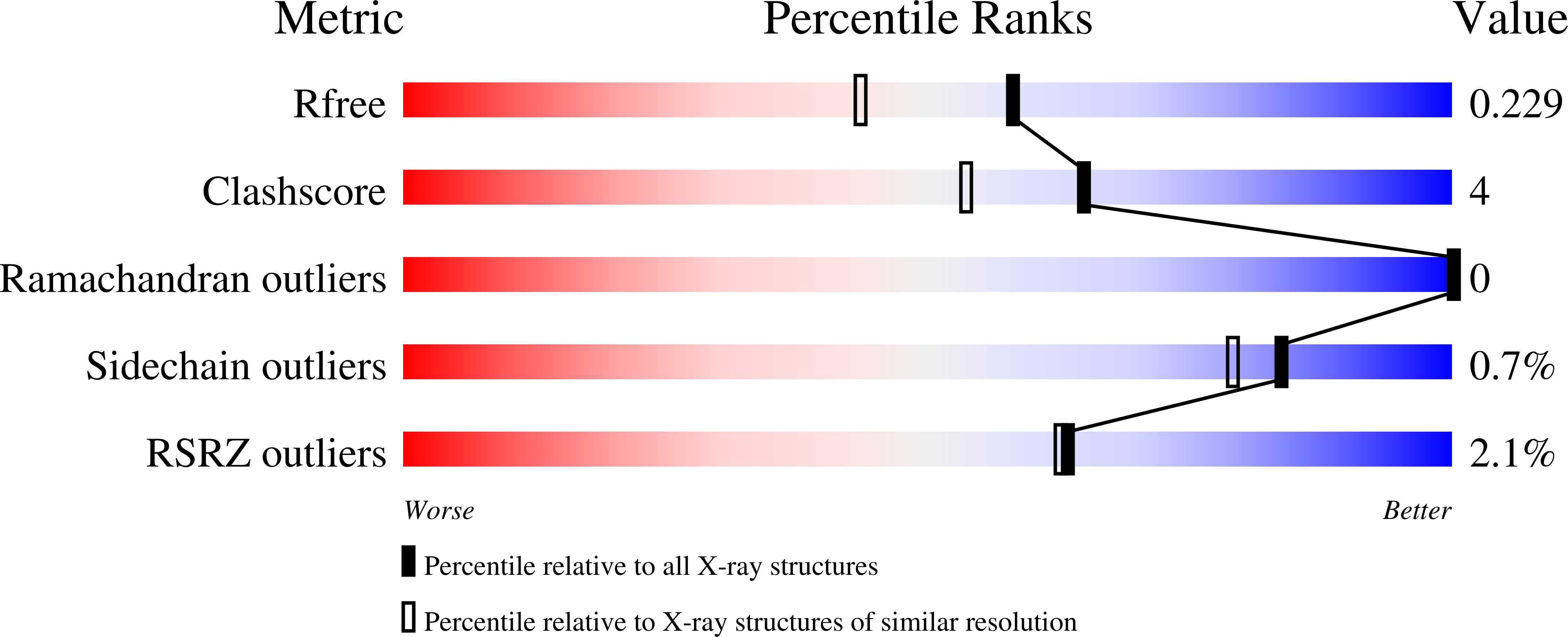
R-Value Free:
0.22
R-Value Work:
0.19
R-Value Observed:
0.19
Space Group:
P 2 21 21