
Deposition Date
2022-09-09
Release Date
2023-06-28
Last Version Date
2024-10-16
Entry Detail
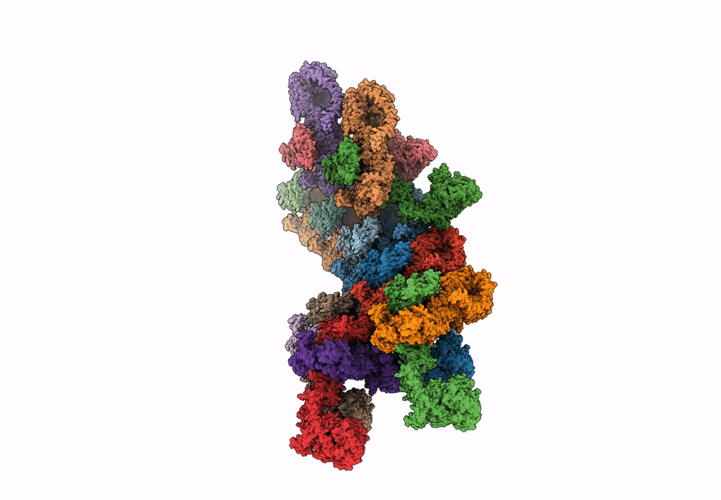
PDB ID:
8EFT
Keywords:
Title:
CryoEM of the soluble OPA1 interfaces from the apo helical assembly on a lipid membrane
Biological Source:
Source Organism(s):
Homo sapiens (Taxon ID: 9606)
Expression System(s):
Method Details:
Experimental Method:
Resolution:
9.68 Å
Aggregation State:
HELICAL ARRAY
Reconstruction Method:
HELICAL
