8EBG image
Deposition Date 2022-08-31
Release Date 2022-09-14
Last Version Date 2024-11-20
Entry Detail
PDB ID:
8EBG
Title:
Crystal structure of the probable FhuD FeIII-dicitrate-binding domain protein FecB from Mycobacterium tuberculosis
Biological Source:
Source Organism:
Method Details:
Experimental Method:
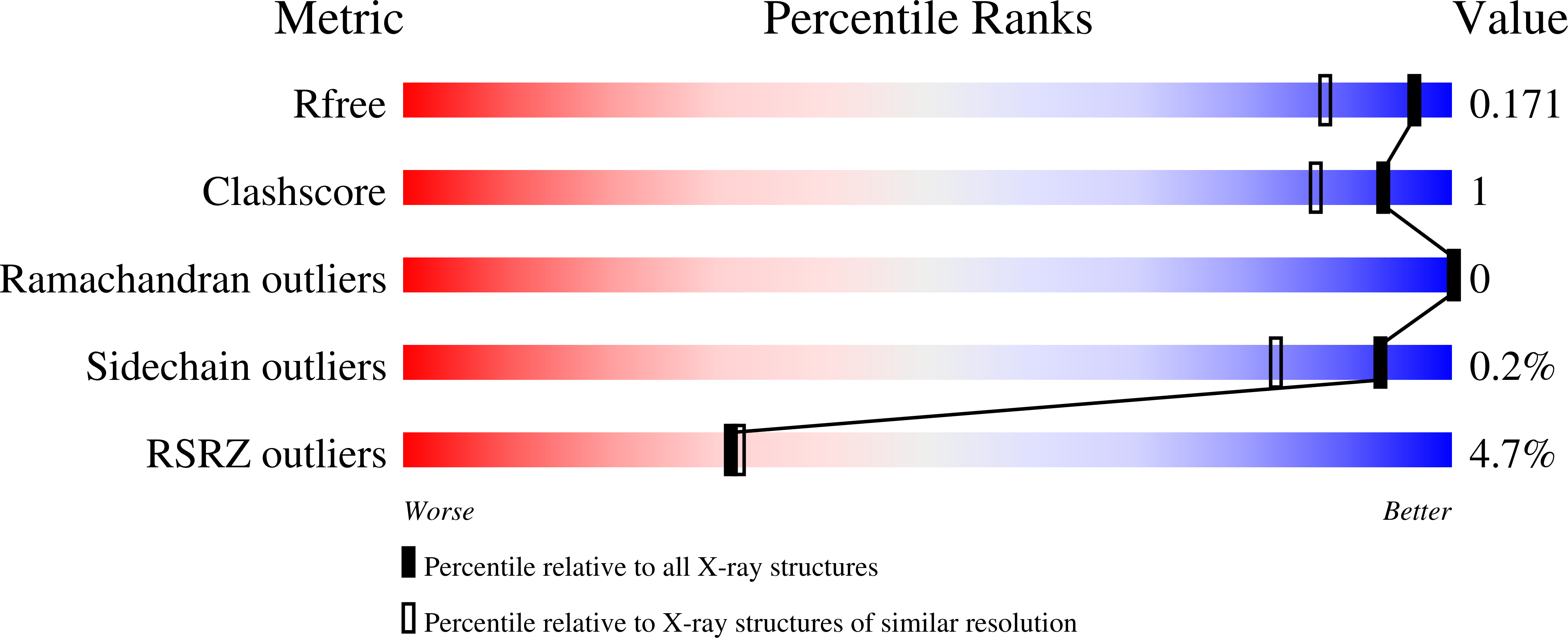
Resolution:
1.43 Å
R-Value Free:
0.17
R-Value Work:
0.14
R-Value Observed:
0.14
Space Group:
P 1 21 1
Macromolecular Entities
Structures with similar UniProt ID
Protein Blast
Polymer Type:polypeptide(L)
Molecule:FEIII-dicitrate-binding periplasmic lipoprotein FecB
Gene (Uniprot):fecB
Chain IDs:A, B
Chain Length:336
Number of Molecules:2
Biological Source:Mycobacterium tuberculosis H37Rv
Modified Residue
Compound ID Chain ID Parent Comp ID Details 2D Image
MSE A MET modified residue
Primary Citation
Crystal structure of the probable FhuD FeIII-dicitrate-binding domain protein FecB from Mycobacterium tuberculosis
To Be Published ? ? ? (?)
Primary Citation of related structures
Feedback Form
Name
Email
Institute
Feedback