Deposition Date
2022-08-17
Release Date
2023-01-11
Last Version Date
2025-06-04
Entry Detail
PDB ID:
8E40
Keywords:
Title:
Full-length APOBEC3G in complex with HIV-1 Vif, CBF-beta, and fork RNA
Biological Source:
Source Organism(s):
Macaca mulatta (Taxon ID: 9544)
Human immunodeficiency virus 1 (Taxon ID: 11676)
Homo sapiens (Taxon ID: 9606)
Escherichia coli (Taxon ID: 562)
Human immunodeficiency virus 1 (Taxon ID: 11676)
Homo sapiens (Taxon ID: 9606)
Escherichia coli (Taxon ID: 562)
Expression System(s):
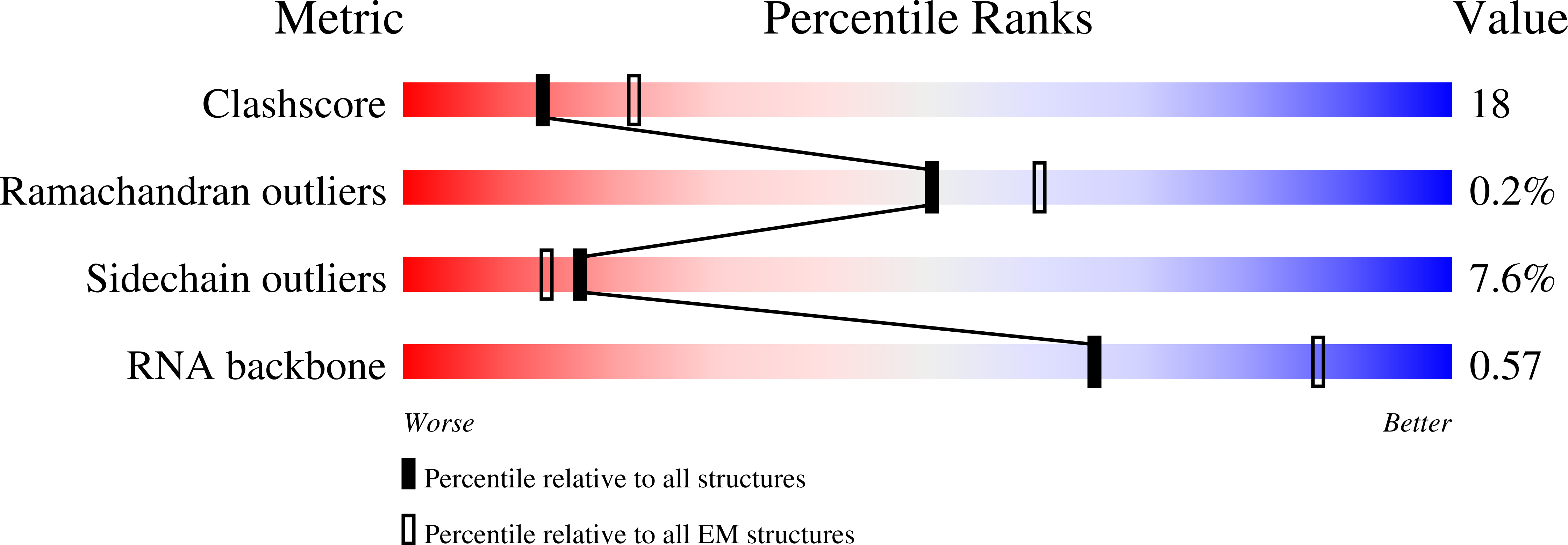
Method Details:
Experimental Method:
Resolution:
3.57 Å
Aggregation State:
PARTICLE
Reconstruction Method:
SINGLE PARTICLE