
Deposition Date
2022-08-09
Release Date
2023-05-03
Last Version Date
2024-11-20
Entry Detail
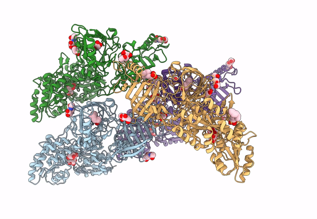
PDB ID:
8E0P
Keywords:
Title:
Crystal structure of mouse APCDD1 in fusion with engineered MBP
Biological Source:
Source Organism:
Escherichia coli (Taxon ID: 562)
Mus musculus (Taxon ID: 10090)
Mus musculus (Taxon ID: 10090)
Host Organism:
Method Details:
Experimental Method:
Resolution:
2.33 Å
R-Value Free:
0.23
R-Value Work:
0.19
R-Value Observed:
0.19
Space Group:
P 1 21 1
