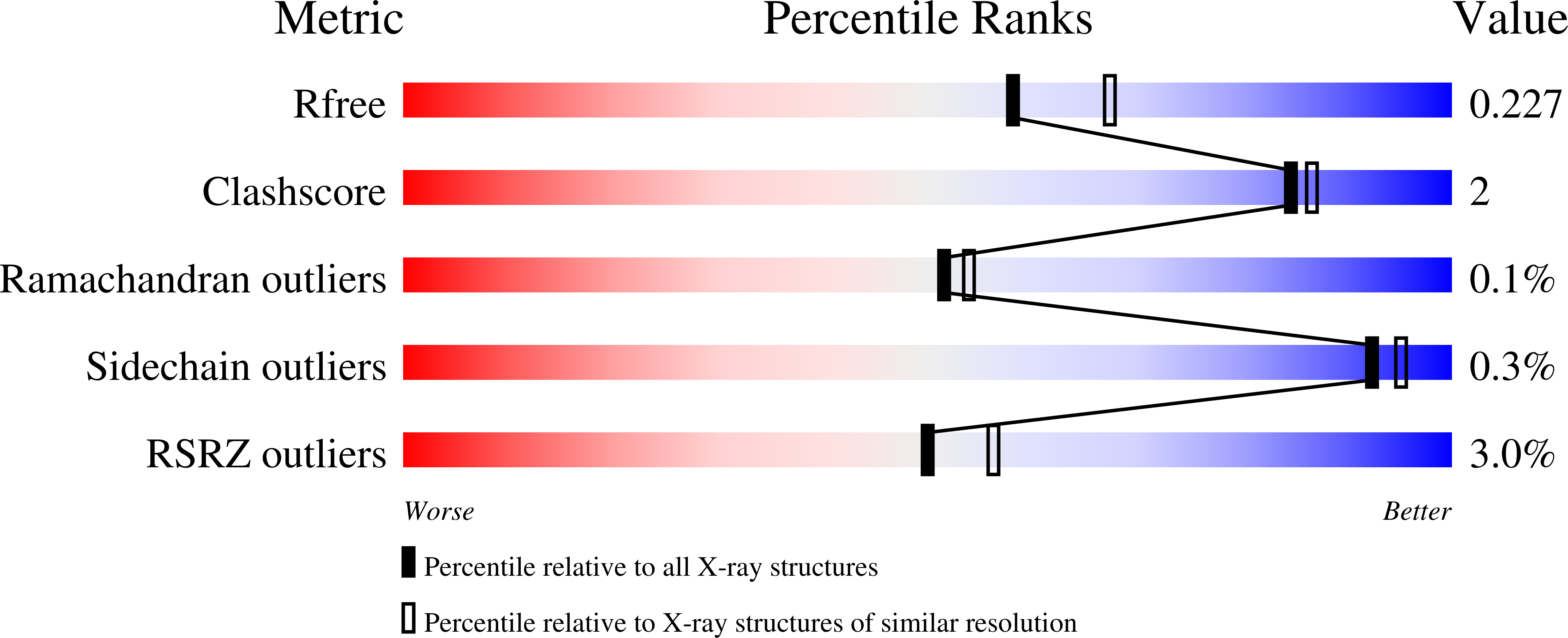
8DWR image
Deposition Date 2022-08-01
Release Date 2023-08-02
Last Version Date 2024-03-13
Entry Detail
PDB ID:
8DWR
Keywords:
Title:
Crystal structure of the L333V variant of catalase-peroxidase from Mycobacterium tuberculosis
Biological Source:
Source Organism:
Host Organism:
Method Details:
Experimental Method:
Resolution:
2.10 Å
R-Value Free:
0.22
R-Value Work:
0.18
R-Value Observed:
0.18
Space Group:
P 1 21 1
Macromolecular Entities
Polymer Type:polypeptide(L)
Molecule:Catalase-peroxidase
Gene (Uniprot):katG
Mutations:L333V
Chain IDs:A, B, C, D
Chain Length:746
Number of Molecules:4
Biological Source:Mycobacterium tuberculosis
Modified Residue
Compound ID Chain ID Parent Comp ID Details 2D Image
TOX A TRP modified residue
Primary Citation
Characterization of a catalase-peroxidase variant (L333V-KatG) identified in an INH-resistant Mycobacterium tuberculosis clinical isolate.
Biochem Biophys Rep 37 101649 101649 (2024)
PMID: 38318524 DOI: 10.1016/j.bbrep.2024.101649

Abstact

Mycobacterium tuberculosis catalase-peroxidase (Mt-KatG) is a bifunctional heme-dependent enzyme that has been shown to activate isoniazid (INH), the widely used antibiotic against tuberculosis (TB). The L333V-KatG variant has been associated with INH resistance in clinical M. tuberculosis isolates from Mexico. To understand better the mechanisms of INH activation, its catalytic properties (catalase, peroxidase, and IN-NAD formation) and crystal structure were compared with those of the wild-type enzyme (WT-KatG). The rate of IN-NAD formation mediated by WT-KatG was 23% greater than L333V-KatG when INH concentration is varied. In contrast to WT-KatG, the crystal structure of the L333V-KatG variant has a perhydroxy modification of the indole nitrogen of W107 from MYW adduct. L333V-KatG shows most of the active site residues in a similar position to WT-KatG; only R418 is in the R-conformation instead of the double R and Y conformation present in WT-KatG. L333V-KatG shows a small displacement respect to WT-KatG in the helix from R385 to L404 towards the mutation site, an increase in length of the coordination bond between H270 and heme Fe, and a longer H-bond between proximal D381 and W321, compared to WT-KatG; these small displacements could explain the altered redox potential of the heme, and result in a less active and stable enzyme.

Legend

Protein

Chemical

Disease

Primary Citation of related structures