8DUF image
Deposition Date 2022-07-27
Release Date 2023-03-15
Last Version Date 2023-10-25
Entry Detail
PDB ID:
8DUF
Keywords:
Title:
Crystal structure of Venezuelan Equine Encephalitis alphavirus (VEEV) nonstructural protein 2 (nsp2) (K741A/K767A) protease domain
Biological Source:
Source Organism:
Host Organism:
Method Details:
Experimental Method:
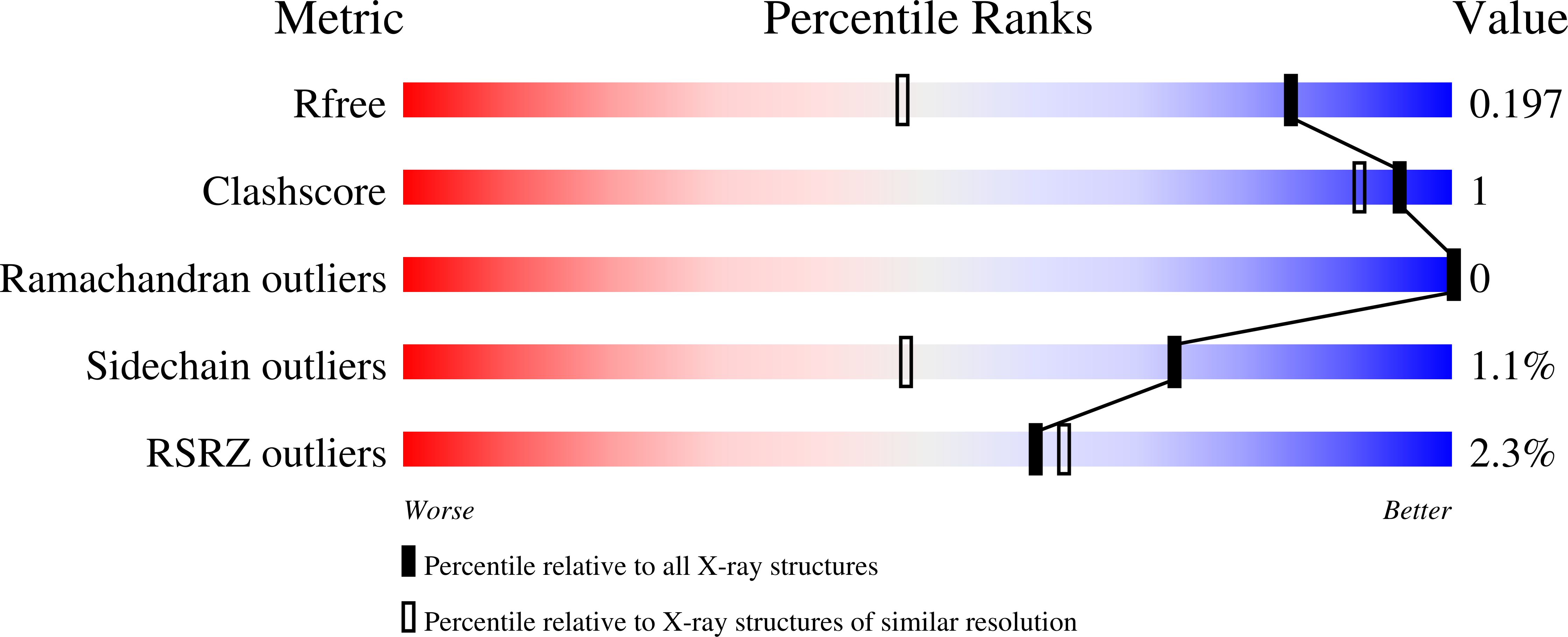
Resolution:
1.46 Å
R-Value Free:
0.19
R-Value Work:
0.15
R-Value Observed:
0.15
Space Group:
P 21 21 21
Macromolecular Entities
Polymer Type:polypeptide(L)
Molecule:nsp2 protease domain
Mutations:K1276A/K1302A
Chain IDs:A
Chain Length:323
Number of Molecules:1
Biological Source:Venezuelan equine encephalitis virus
Primary Citation
Self-inhibited State of Venezuelan Equine Encephalitis Virus (VEEV) nsP2 Cysteine Protease: A Crystallographic and Molecular Dynamics Analysis.
J.Mol.Biol. 435 168012 168012 (2023)
PMID: 36792007 DOI: 10.1016/j.jmb.2023.168012

Abstact

The Venezuelan equine encephalitis virus (VEEV) belongs to the Togaviridae family and is pathogenic to both humans and equines. The VEEV non-structural protein 2 (nsP2) is a cysteine protease (nsP2pro) that processes the polyprotein and thus it is a drug target for inhibitor discovery. The atomic structure of the VEEV nsP2 catalytic domain was previously characterized by both X-ray crystallography and computational studies. A modified nsP2pro harboring a N475A mutation in the N terminus was observed to exhibit an unexpected conformation: the N-terminal residues bind to the active site, mimicking binding of a substrate. The large conformational change of the N terminus was assumed to be induced by the N475A mutation, as N475 has an important role in stabilization of the N terminus and the active site. This conformation was first observed in the N475A mutant, but we also found it while determining a crystal structure of the catalytically active nsP2pro containing the wild-type N475 active site residue and K741A/K767A surface entropy reduction mutations. This suggests that the N475A mutation is not a prerequisite for self-inhibition. Here, we describe a high resolution (1.46 Å) crystal structure of a truncated nsP2pro (residues 463-785, K741A/K767A) and analyze the structure further by molecular dynamics to study the active and self-inhibited conformations of nsP2pro and its N475A mutant. A comparison of the different conformations of the N-terminal residues sheds a light on the interactions that play an important role in the stabilization of the enzyme.

Legend

Protein

Chemical

Disease

Primary Citation of related structures