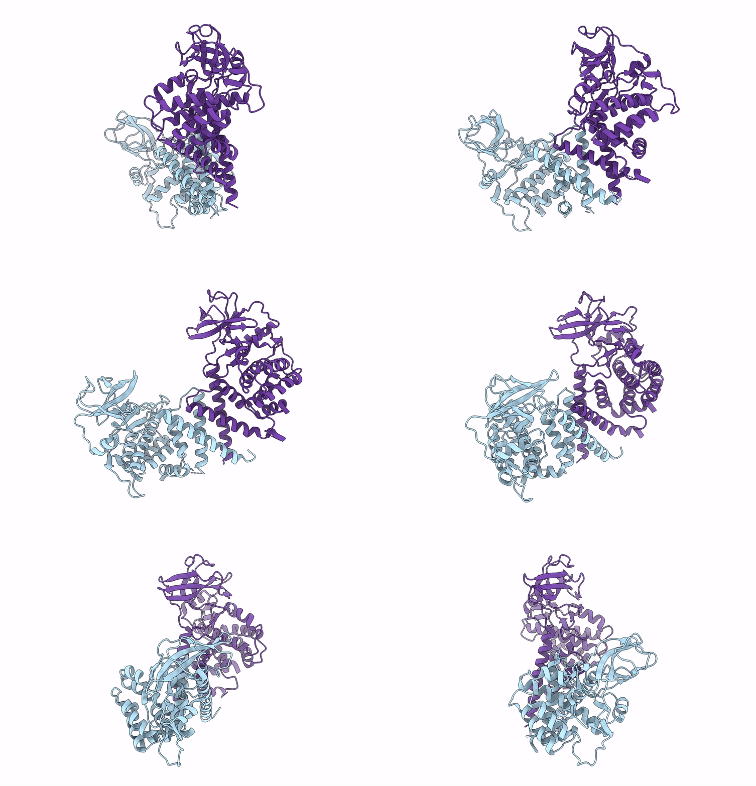
8DS6 image
Deposition Date 2022-07-21
Release Date 2023-06-28
Last Version Date 2024-06-12
Entry Detail
PDB ID:
8DS6
Title:
Structure of the PEAK3 pseudokinase homodimer
Biological Source:
Source Organism:
Homo sapiens (Taxon ID: 9606)
Host Organism:
Method Details:
Experimental Method:
Resolution:
4.90 Å
Aggregation State:
PARTICLE
Reconstruction Method:
SINGLE PARTICLE
Macromolecular Entities
Polymer Type:polypeptide(L)
Molecule:Protein PEAK3
Gene (Uniprot):PEAK3
Chain IDs:A, B
Chain Length:491
Number of Molecules:2
Biological Source:Homo sapiens
Ligand Molecules
Primary Citation
Structural insights into regulation of the PEAK3 pseudokinase scaffold by 14-3-3.
Nat Commun 14 3543 3543 (2023)
PMID: 37336883 DOI: 10.1038/s41467-023-38864-0

Abstact

PEAK pseudokinases are molecular scaffolds which dimerize to regulate cell migration, morphology, and proliferation, as well as cancer progression. The mechanistic role dimerization plays in PEAK scaffolding remains unclear, as there are no structures of PEAKs in complex with their interactors. Here, we report the cryo-EM structure of dimeric PEAK3 in complex with an endogenous 14-3-3 heterodimer. Our structure reveals an asymmetric binding mode between PEAK3 and 14-3-3 stabilized by one pseudokinase domain and the SHED domain of the PEAK3 dimer. The binding interface contains a canonical phosphosite-dependent primary interaction and a unique secondary interaction not observed in previous structures of 14-3-3/client complexes. Additionally, we show that PKD regulates PEAK3/14-3-3 binding, which when prevented leads to PEAK3 nuclear enrichment and distinct protein-protein interactions. Altogether, our data demonstrate that PEAK3 dimerization forms an unusual secondary interface for 14-3-3 binding, facilitating 14-3-3 regulation of PEAK3 localization and interactome diversity.

Legend

Protein

Chemical

Disease

Primary Citation of related structures