Deposition Date
2022-07-18
Release Date
2024-01-17
Last Version Date
2024-01-17
Entry Detail
PDB ID:
8DQD
Keywords:
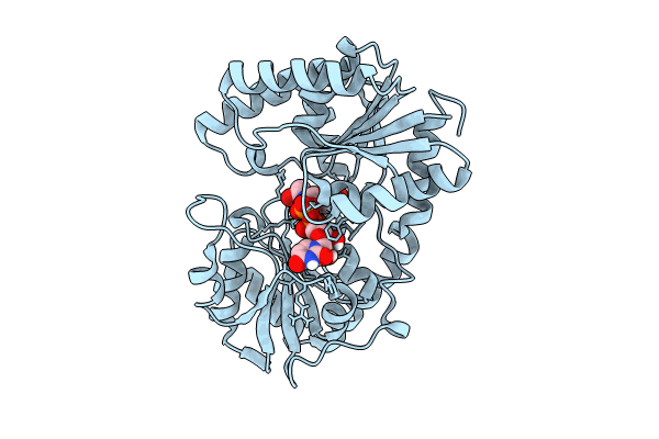
Title:
Structure of the Campylobacter concisus glycosyltransferase PglA
Biological Source:
Source Organism(s):
Campylobacter concisus (Taxon ID: 199)
Expression System(s):
Method Details:
Experimental Method:
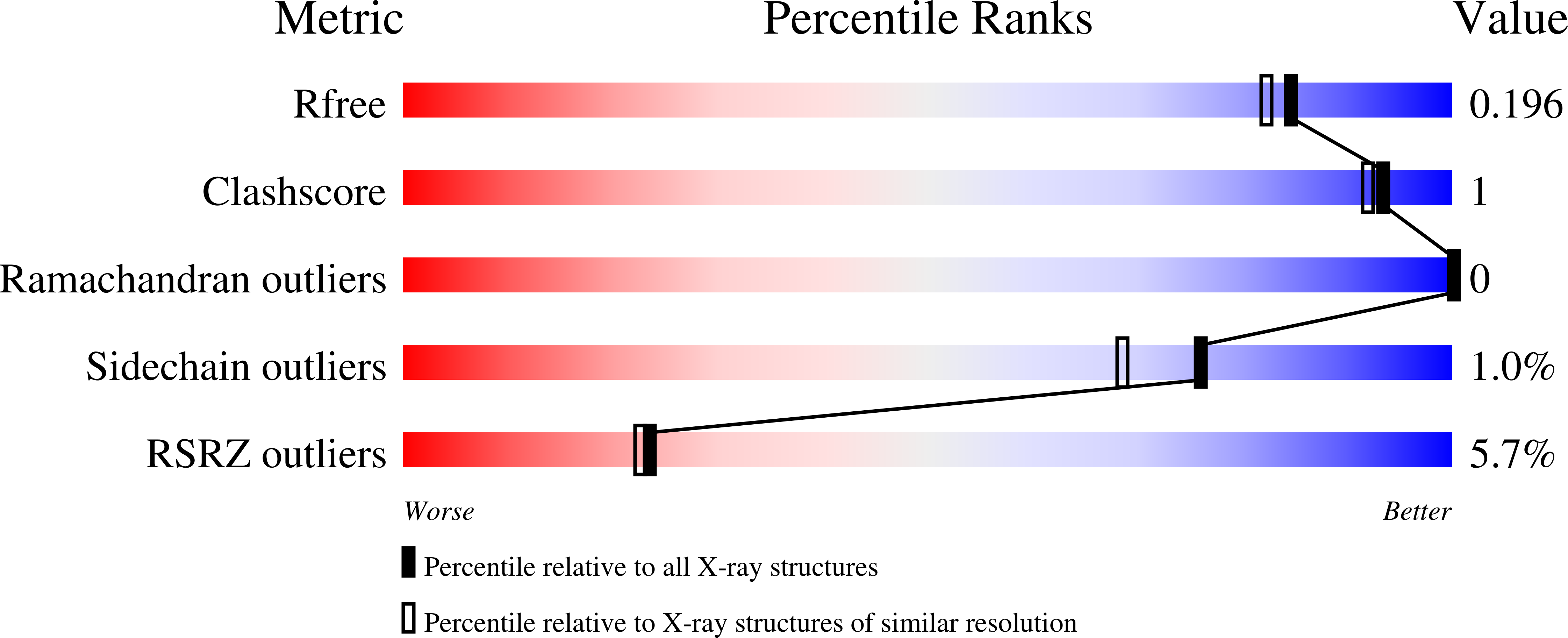
Resolution:
1.78 Å
R-Value Free:
0.19
R-Value Work:
0.17
R-Value Observed:
0.17
Space Group:
P 31 2 1