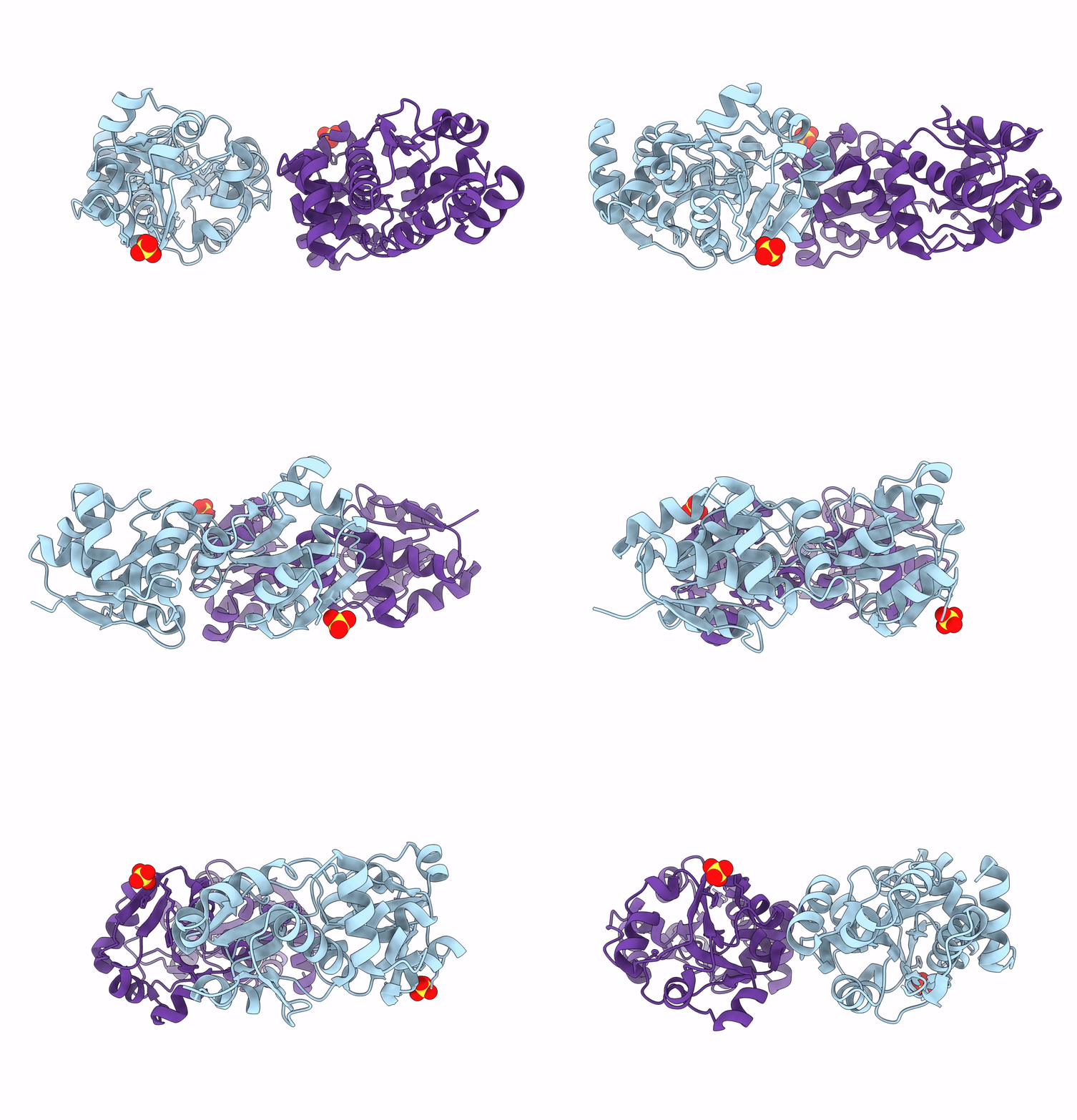
8DP6 image
Entry Detail
PDB ID:
8DP6
Title:
Crystal structure of Helicobacter pylori EgtU
Biological Source:
Host Organism:
PDB Version:
Deposition Date:
2022-07-15
Release Date:
2022-11-09
Method Details:
Experimental Method:
Resolution:
1.30 Å
R-Value Free:
0.18
R-Value Work:
0.17
Space Group:
C 1 2 1
Macromolecular Entities
Polymer Type:polypeptide(L)
Description:Osmoprotection protein
Chain IDs:A, B
Chain Length:272
Number of Molecules:2
Biological Source:Helicobacter pylori (strain P12)
Ligand Molecules
Primary Citation
A microbial transporter of the dietary antioxidant ergothioneine.
Cell 185 4526 4540.e18 (2022)
PMID: 36347253 DOI: 10.1016/j.cell.2022.10.008

Abstact

Low-molecular-weight (LMW) thiols are small-molecule antioxidants required for the maintenance of intracellular redox homeostasis. However, many host-associated microbes, including the gastric pathogen Helicobacter pylori, unexpectedly lack LMW-thiol biosynthetic pathways. Using reactivity-guided metabolomics, we identified the unusual LMW thiol ergothioneine (EGT) in H. pylori. Dietary EGT accumulates to millimolar levels in human tissues and has been broadly implicated in mitigating disease risk. Although certain microorganisms synthesize EGT, we discovered that H. pylori acquires this LMW thiol from the host environment using a highly selective ATP-binding cassette transporter-EgtUV. EgtUV confers a competitive colonization advantage in vivo and is widely conserved in gastrointestinal microbes. Furthermore, we found that human fecal bacteria metabolize EGT, which may contribute to production of the disease-associated metabolite trimethylamine N-oxide. Collectively, our findings illustrate a previously unappreciated mechanism of microbial redox regulation in the gut and suggest that inter-kingdom competition for dietary EGT may broadly impact human health.

Legend

Protein

Chemical

Disease

Primary Citation of related structures