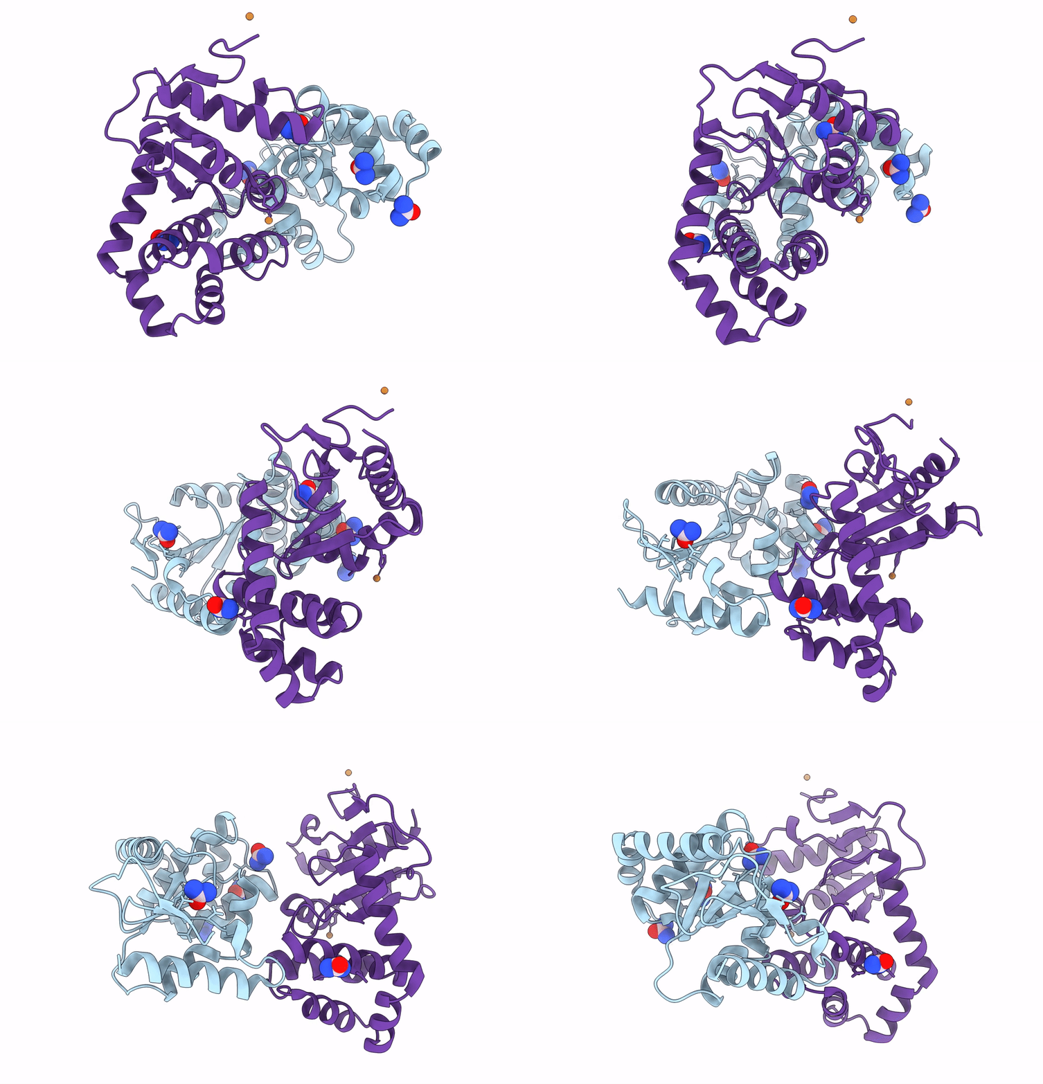
8DG0 image
Entry Detail
PDB ID:
8DG0
Keywords:
Title:
Crystal Structure of EcDsbA in a complex with Urea
Biological Source:
Source Organism:
PDB Version:
Deposition Date:
2022-06-23
Release Date:
2022-12-07
Method Details:
Experimental Method:
Resolution:
2.50 Å
R-Value Free:
0.25
R-Value Work:
0.20
R-Value Observed:
0.20
Space Group:
C 1 2 1
Macromolecular Entities
Polymer Type:polypeptide(L)
Description:Thiol:disulfide interchange protein DsbA
Chain IDs:A, B
Chain Length:189
Number of Molecules:2
Biological Source:Escherichia coli K-12
Primary Citation
Fragment screening libraries for the identification of protein hot spots and their minimal binding pharmacophores.
Rsc Med Chem 14 135 143 (2023)
PMID: 36760747 DOI: 10.1039/d2md00253a

Abstact

Fragment-based drug design relies heavily on structural information for the elaboration and optimisation of hits. The ability to identify neighbouring binding hot spots, energetically favourable interactions and conserved binding motifs in protein structures through X-ray crystallography can inform the evolution of fragments into lead-like compounds through structure-based design. The composition of fragment libraries can be designed and curated to fit this purpose and herein, we describe and compare screening libraries containing compounds comprising between 2 and 18 heavy atoms. We evaluate the properties of the compounds in these libraries and assess their ability to probe protein surfaces for binding hot spots.

Legend

Protein

Chemical

Disease

Primary Citation of related structures