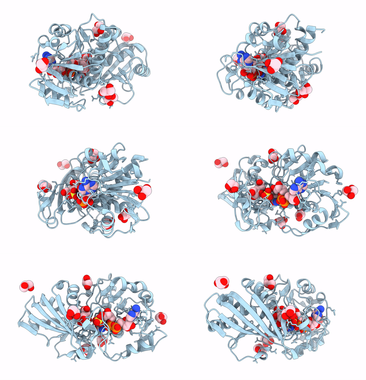
8DE5 image
Deposition Date 2022-06-19
Release Date 2023-06-21
Last Version Date 2024-11-13
Entry Detail
PDB ID:
8DE5
Keywords:
Title:
Structure of glyceraldehyde-3-phosphate dehydrogenase from Paracoccidioides lutzii
Biological Source:
Source Organism:
Host Organism:
Method Details:
Experimental Method:
Resolution:
2.02 Å
R-Value Free:
0.19
R-Value Work:
0.16
R-Value Observed:
0.16
Space Group:
I 41 2 2
Macromolecular Entities
Structures with similar UniProt ID
Protein Blast
Polymer Type:polypeptide(L)
Molecule:Glyceraldehyde-3-phosphate dehydrogenase
Gene (Uniprot):GPD
Chain IDs:A
Chain Length:338
Number of Molecules:1
Biological Source:Paracoccidioides lutzii Pb01
Modified Residue
Compound ID Chain ID Parent Comp ID Details 2D Image
CSD A CYS modified residue
Primary Citation
Structure of glyceraldehyde-3-phosphate dehydrogenase from Paracoccidioides lutzii in complex with an aldonic sugar acid.
Biochimie 218 20 33 (2023)
PMID: 37709188 DOI: 10.1016/j.biochi.2023.09.013

Abstact

The pathogen Paracoccidioides lutzii (Pb01) is found in South America countries Colombia, Ecuador, Venezuela and Brazil, especially in the central, west, and north regions of the latter. It belongs to the Ajellomycetaceae family, Onygenales order, and is typically thermodimorphic, presenting yeast cells when it grows in animal tissues, but mycelia when in the environment, where it produces the infectious propagule. This fungus is one of the etiologic agents of Paracoccidioidomycosis (PCM), the most important endemic fungal infection in Latin America. Investigations on its genome have contributed to a better understanding about its metabolism and revealed the complexity of several metabolic glycolytic pathways. Glyceraldehyde-3-Phosphate Dehydrogenase from Paracoccidioides lutzii (PlGAPDH) is considered a moonlighting protein and participates in several biological processes of this pathogen. The enzyme was expressed and purified, as seen in SDS-PAGE gel, crystallized and had its three dimensional structure (3D) determined in complex with NAD+, a sulphate ion and d-galactonic acid, therefore, a type of 'GAA site'. It is the first GAPDH structure to show this chemical type in this site and how this protein can bind an acid derived from oxidation of a linear hexose.

Legend

Protein

Chemical

Disease

Primary Citation of related structures