Deposition Date
2022-06-07
Release Date
2023-06-14
Last Version Date
2024-10-23
Entry Detail
PDB ID:
8D7K
Keywords:
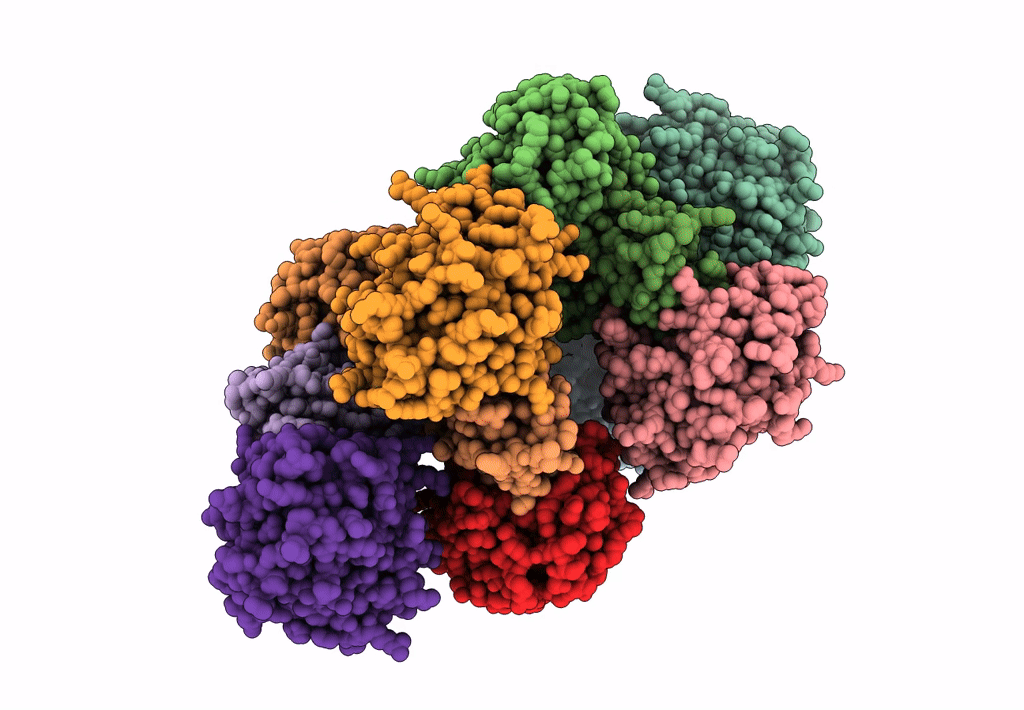
Title:
Bifunctional Inhibition of Neutrophil Elastase and Cathepsin G by Eap2 from S. aureus
Biological Source:
Source Organism(s):
Staphylococcus aureus subsp. aureus (Taxon ID: 158878)
Homo sapiens (Taxon ID: 9606)
Homo sapiens (Taxon ID: 9606)
Expression System(s):
Method Details:
Experimental Method:
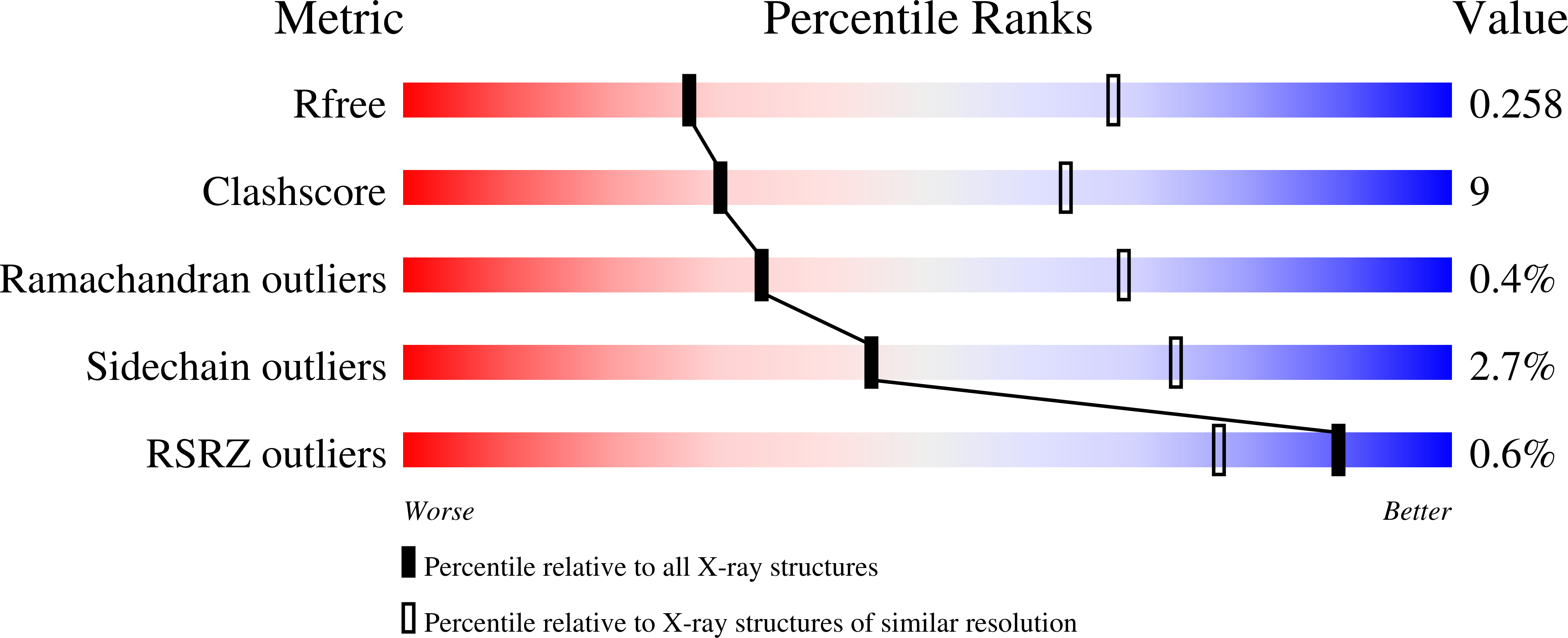
Resolution:
3.10 Å
R-Value Free:
0.25
R-Value Work:
0.18
R-Value Observed:
0.18
Space Group:
P 1 21 1