8D4X image
Deposition Date 2022-06-02
Release Date 2023-04-19
Last Version Date 2025-05-28
Entry Detail
PDB ID:
8D4X
Keywords:
Title:
Structure of the human UBR5 HECT-type E3 ubiquitin ligase in a dimeric form
Biological Source:
Source Organism:
Homo sapiens (Taxon ID: 9606)
Method Details:
Experimental Method:
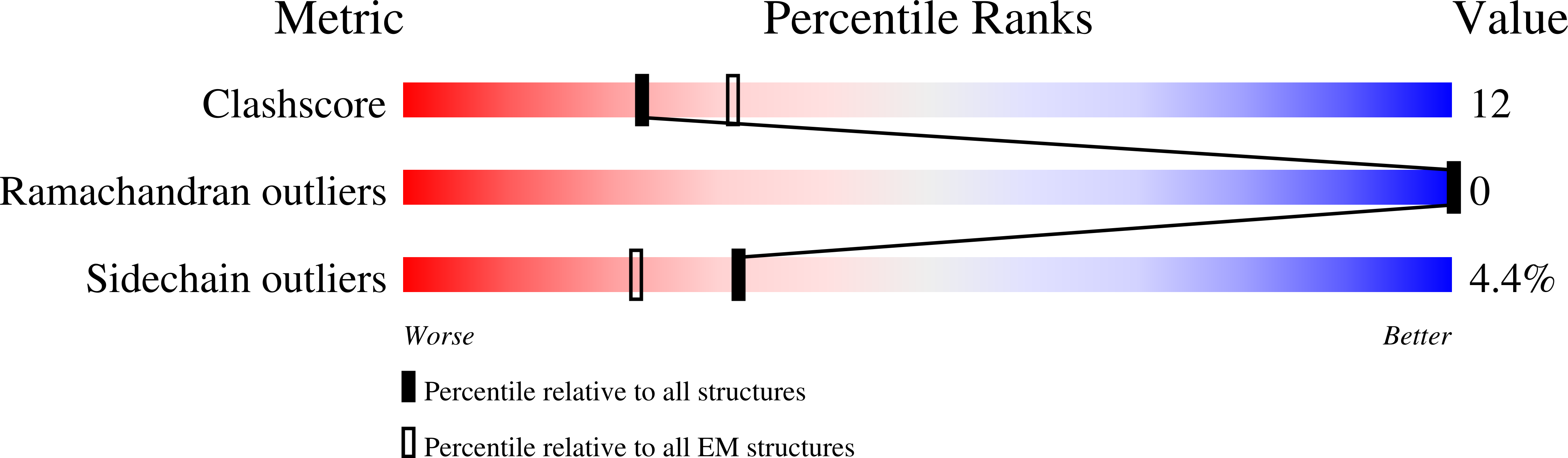
Resolution:
2.80 Å
Aggregation State:
PARTICLE
Reconstruction Method:
SINGLE PARTICLE
Macromolecular Entities
Structures with similar UniProt ID
Protein Blast
Polymer Type:polypeptide(L)
Molecule:E3 ubiquitin-protein ligase UBR5
Gene (Uniprot):UBR5
Chain IDs:A, B
Chain Length:2806
Number of Molecules:2
Biological Source:Homo sapiens
Ligand Molecules
Primary Citation
Structure of the human UBR5 E3 ubiquitin ligase.
Structure 31 541 552.e4 (2023)
PMID: 37040767 DOI: 10.1016/j.str.2023.03.010

Abstact

The human UBR5 is a single polypeptide chain homology to E6AP C terminus (HECT)-type E3 ubiquitin ligase essential for embryonic development in mammals. Dysregulated UBR5 functions like an oncoprotein to promote cancer growth and metastasis. Here, we report that UBR5 assembles into a dimer and a tetramer. Our cryoelectron microscopy (cryo-EM) structures reveal that two crescent-shaped UBR5 monomers assemble head to tail to form the dimer, and two dimers bind face to face to form the cage-like tetramer with all four catalytic HECT domains facing the central cavity. Importantly, the N-terminal region of one subunit and the HECT of the other form an "intermolecular jaw" in the dimer. We show the jaw-lining residues are important for function, suggesting that the intermolecular jaw functions to recruit ubiquitin-loaded E2 to UBR5. Further work is needed to understand how oligomerization regulates UBR5 ligase activity. This work provides a framework for structure-based anticancer drug development and contributes to a growing appreciation of E3 ligase diversity.

Legend

Protein

Chemical

Disease

Primary Citation of related structures
Feedback Form
Name
Email
Institute
Feedback