Deposition Date
2022-06-02
Release Date
2022-12-21
Last Version Date
2024-11-13
Entry Detail
PDB ID:
8D4S
Keywords:
Title:
Crystal Structure of Cathepsin G Inhibited by Eap1 from S. aureus
Biological Source:
Source Organism(s):
Staphylococcus aureus subsp. aureus Mu50 (Taxon ID: 158878)
Homo sapiens (Taxon ID: 9606)
Homo sapiens (Taxon ID: 9606)
Expression System(s):
Method Details:
Experimental Method:
Resolution:
1.95 Å
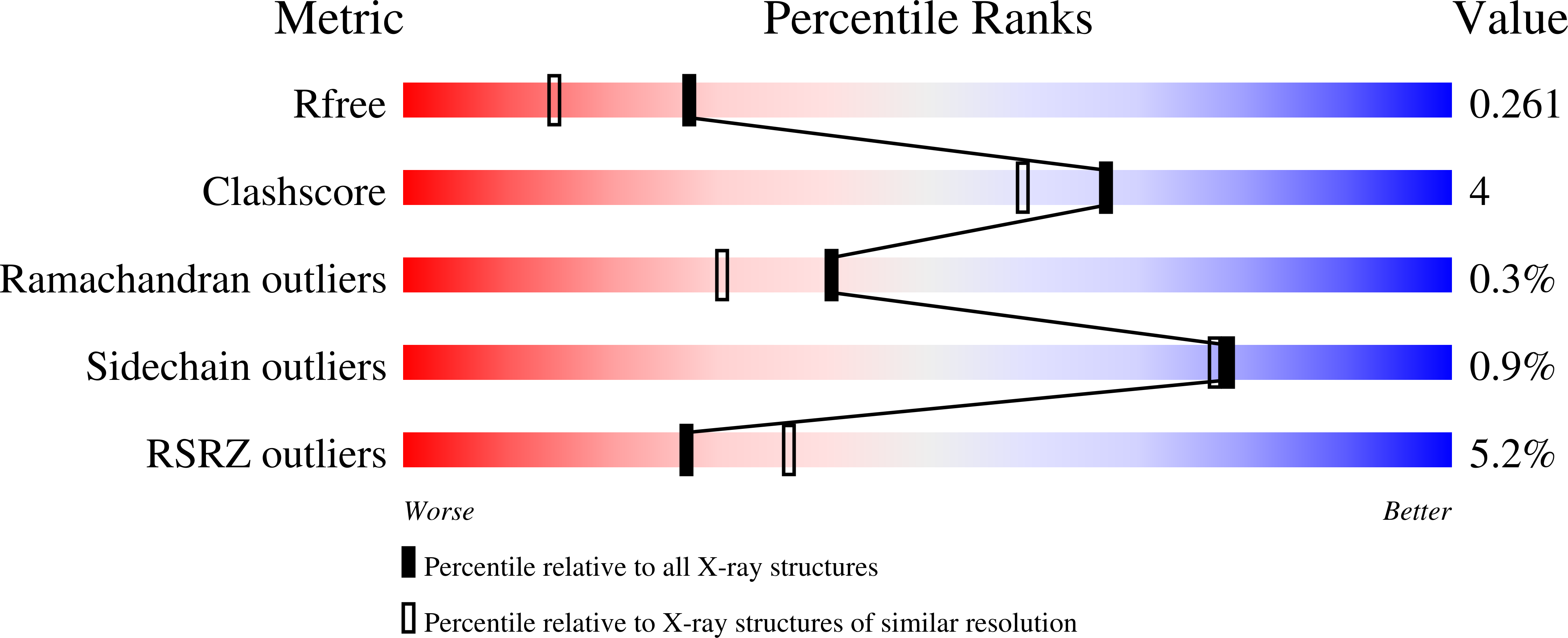
R-Value Free:
0.26
R-Value Work:
0.20
R-Value Observed:
0.21
Space Group:
F 2 2 2