8CXO image
Deposition Date 2022-05-22
Release Date 2022-08-03
Last Version Date 2024-10-09
Entry Detail
PDB ID:
8CXO
Title:
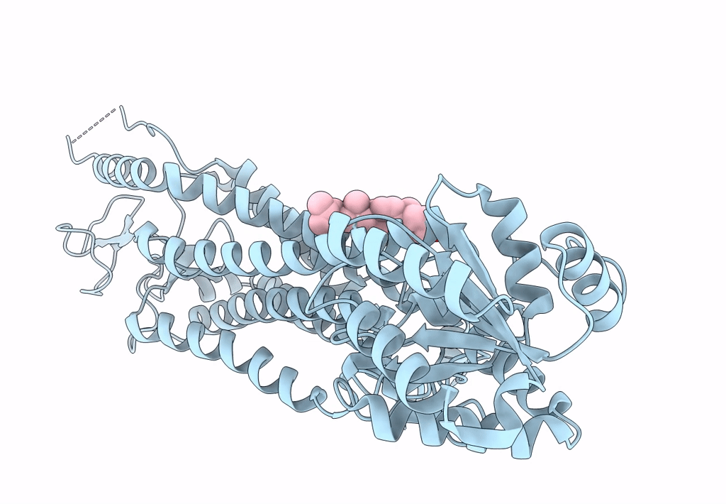
Cryo-EM structure of the unliganded mSMO-PGS2 in a lipidic environment
Biological Source:
Source Organism:
Host Organism:
Method Details:
Experimental Method:
Resolution:
3.70 Å
Aggregation State:
PARTICLE
Reconstruction Method:
SINGLE PARTICLE
Macromolecular Entities
Polymer Type:polypeptide(L)
Molecule:Smoothened homolog, GlgA glycogen synthase chimera
Gene (Uniprot):Smo, PAB2292
Chain IDs:A
Chain Length:719
Number of Molecules:1
Biological Source:Mus musculus, Pyrococcus abyssi (strain GE5 / Orsay)
Ligand Molecules
Primary Citation
Fusion protein strategies for cryo-EM study of G protein-coupled receptors.
Nat Commun 13 4366 4366 (2022)
PMID: 35902590 DOI: 10.1038/s41467-022-32125-2

Abstact

Single particle cryogenic-electron microscopy (cryo-EM) is used extensively to determine structures of activated G protein-coupled receptors (GPCRs) in complex with G proteins or arrestins. However, applying it to GPCRs without signaling proteins remains challenging because most receptors lack structural features in their soluble domains to facilitate image alignment. In GPCR crystallography, inserting a fusion protein between transmembrane helices 5 and 6 is a highly successful strategy for crystallization. Although a similar strategy has the potential to broadly facilitate cryo-EM structure determination of GPCRs alone without signaling protein, the critical determinants that make this approach successful are not yet clear. Here, we address this shortcoming by exploring different fusion protein designs, which lead to structures of antagonist bound A2A adenosine receptor at 3.4 Å resolution and unliganded Smoothened at 3.7 Å resolution. The fusion strategies explored here are likely applicable to cryo-EM interrogation of other GPCRs and small integral membrane proteins.

Legend

Protein

Chemical

Disease

Primary Citation of related structures