Deposition Date
2022-05-19
Release Date
2022-09-28
Last Version Date
2023-10-25
Entry Detail
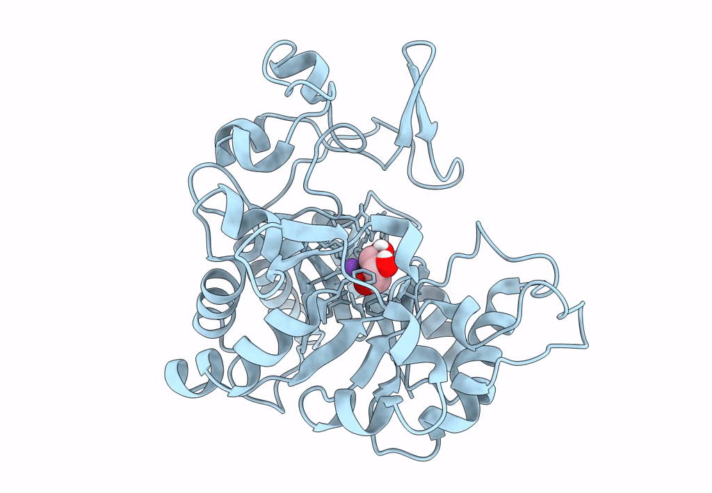
PDB ID:
8CWP
Keywords:
Title:
X-ray crystal structure of NTHi Protein D bound to a putative glycerol moiety
Biological Source:
Source Organism(s):
Haemophilus influenzae (Taxon ID: 727)
Expression System(s):
Method Details:
Experimental Method:
Resolution:
1.80 Å
R-Value Free:
0.18
R-Value Work:
0.15
R-Value Observed:
0.15
Space Group:
P 43 21 2