8CQE image
Entry Detail
PDB ID:
8CQE
Keywords:
Title:
pVHL:EloB:EloC in complex with (2S,4R)-1-((S)-2-(1-Fluorocyclopropane-1-carboxamido)-3,3-dimethylbutanoyl)-4-hydroxy-N-((S)-1-(2-methyl-4-(4-methylthiazol-5-yl)phenyl)ethyl)pyrrolidine-2-carboxamide (Compound 37)
Biological Source:
Source Organism:
PDB Version:
Deposition Date:
2023-03-06
Release Date:
2023-09-27
Method Details:
Experimental Method:
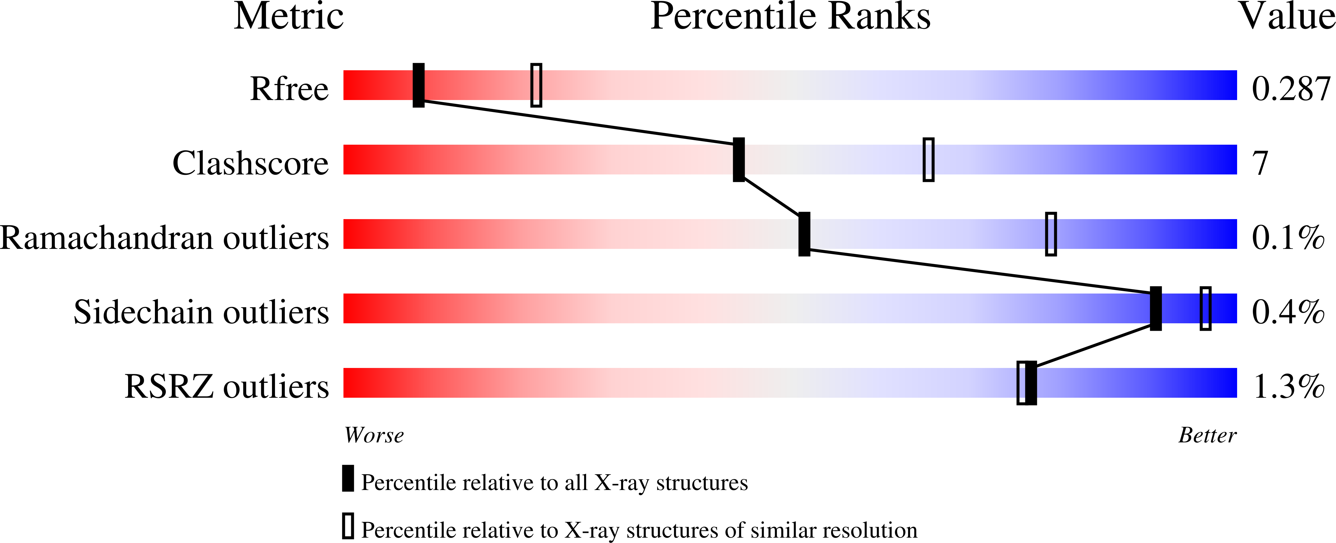
Resolution:
2.85 Å
R-Value Free:
0.29
R-Value Work:
0.24
R-Value Observed:
0.24
Space Group:
P 41 21 2
Macromolecular Entities
Polymer Type:polypeptide(L)
Description:Elongin-B
Chain IDs:A, D, G, J
Chain Length:104
Number of Molecules:4
Biological Source:Homo sapiens
Polymer Type:polypeptide(L)
Description:Elongin-C
Chain IDs:B, E, H, K
Chain Length:97
Number of Molecules:4
Biological Source:Homo sapiens
Polymer Type:polypeptide(L)
Description:von Hippel-Lindau disease tumor suppressor
Chain IDs:C, F, I, L
Chain Length:162
Number of Molecules:4
Biological Source:Homo sapiens
Modified Residue
Compound ID Chain ID Parent Comp ID Details 2D Image
CAS A CYS modified residue
Ligand Molecules
Primary Citation
Expanding the Structural Diversity at the Phenylene Core of Ligands for the von Hippel-Lindau E3 Ubiquitin Ligase: Development of Highly Potent Hypoxia-Inducible Factor-1 alpha Stabilizers.
J.Med.Chem. 66 12776 12811 (2023)
PMID: 37708384 DOI: 10.1021/acs.jmedchem.3c00434

Abstact

Hypoxia-inducible factor-1α (HIF-1α) constitutes the principal mediator of cellular adaptation to hypoxia in humans. The HIF-1α protein level and activity are tightly regulated by the ubiquitin E3 ligase von Hippel-Lindau (VHL). Here, we performed a structure-guided and bioactivity-driven design of new VHL inhibitors. Our iterative and combinatorial strategy focused on chemical variability at the phenylene unit and encompassed further points of diversity. The exploitation of tailored phenylene fragments and the stereoselective installation of the benzylic methyl group provided potent VHL ligands. Three high-resolution structures of VHL-ligand complexes were determined, and bioactive conformations of these ligands were explored. The most potent inhibitor (30) exhibited dissociation constants lower than 40 nM, independently determined by fluorescence polarization and surface plasmon resonance and an enhanced cellular potency, as evidenced by its superior ability to induce HIF-1α transcriptional activity. Our work is anticipated to inspire future efforts toward HIF-1α stabilizers and new ligands for proteolysis-targeting chimera (PROTAC) degraders.

Legend

Protein

Chemical

Disease

Primary Citation of related structures